|
A 29-year-old second gravida mother vaginally
delivered a 1700 g (SFD, Ponderal index <2.1) female child at full
term after an apparently uneventful antenatal period. Examination
revealed craniofacial anomalies including microcephaly, malar hypoplasia,
micrognathia, thin small pointed nose with hypoplasia of the cartilage
(becoming parrot like), narrow and high arched palate, thin and light
hair with hypotrichosis, especially of the eyebrows and eyelashes, and
low set ears (Fig. 1).
The neonate also had bilateral central cataracts and an inspiratory
stridor due to laryngomalacia. Radiological features included platybasia,
shallow sella turcica and absence of the mandibular condyles. All these
features were consistent with dyscephalia mandibulo-oculo-facialis.
The syndrome was described indepen-dently by
Hallermann in 1948 and Streiff in 1950. The narrow upper airway
associated with the craniofacial configuration can lead to feeding and
respiratory problems during early infancy as was seen in our case.
Respiratory infections may contribute to the cause of death. Growth
failure occurs in 50% of the cases and mental retardation is seen in
about 15%. The major handicap is the ocular defect, which usually
culminates in blindness despite surgery.
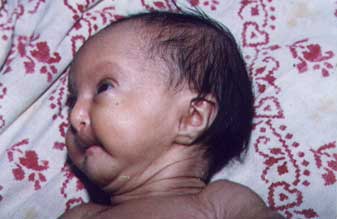
Fig.1. Neonate with small
nose, malar hypoplasia, hypotrichosis and micrognathia
M. Issaivanan,
Verinderjit S. Virdi,
Department of Pediatrics,
Government Medical College and Hospital,
Sector-32, Chandigarh 160 047, India.
|
