1.gif (1892 bytes)                     

Correspondence

Indian Pediatrics 2008; 45:865-866

Congenital Erythropoietic Porphyria


Congenital erythropoietic porphyria (CEP) is a rare cutaneous pophyria which presents with photosensitivity, complicated by presence of hemolytic anemia. There is no permanent cure and the treatment is supportive. A 3 day-old male baby was admitted with neonatal jaundice for phototherapy. The baby developed bullous lesions over both the feet within minutes of starting phototherapy(Fig.1). These lesions were prominently present over the toes and were filled with clear fluid. We observed the color of the urine to be pink. The baby had no hepatosplenomegaly or any evidence of hemolysis. A urine analysis showed the presence of uroporphyrin and coproporphyrin which are not usually detectable suggesting the diagnosis of CEP. A quantitative assay could not be done because of financial reasons. The lesions healed in 2 weeks time following which the patient was discharged with advice regarding photoprotection and regular follow up. The patient is now 3 months old and has not had any further episodes of blister formation.

Fig.1 Edematous lower limbs with scars of surgery. Note also the swelling of the interphalangeal joints of the great toes.

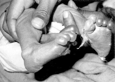
CEP, also known as Gunther’s disease, is an autosomal recessive inherited deficiency of the uroporphyrinogen III cosynthetase enzyme leading to accumulation of type I porphyrins. Less than a hundred cases are reported worldwide(1). The earliest sign of CEP could be brownish discoloration of amniotic fluid or pink to brown staining of the nappies. Severe photosensitivity often begins in the neonatal period itself with blisters developing on exposure to light.There have been earlier case reports of CEP in which the newborns developed photosensitivity following phototherapy but in most of these newborns hemolytic anemia was the main presenting feature(2,3). There are reports of older children and adults with CEP from India also(4,5). Genetic counseling is important for the parents of an affected offspring. Antenatal diagnosis can be made by measuring the uroporphyrin I concentration in the amniotic fluid which is increased as early as 16 weeks in utero.

Diagnosis is made by demonstrating the presence of uroporphyrin and coproporphyrin in the urine and blood. A plasma spectrofluorimetry is seen at 615-620 nm. A positive test with a characteristic history is highly suggestive, although quantitative screening using spectrophotometric or fluorimetric techniques is ideally the best. As this facility was not available to us and the patient was unable to afford it we were not able to do conduct a definitive diagnostic test. We were also not able to do a Woods lamp examination of the eyes or urine because of lack of the facility.

DK Singh,
Ruchi Rai,

Department of Pediatrics,
S N Children Hospital,
M L N Medical College,
Allahabad, UP, India.
E-mail: [email protected]

References


1. Sarkany RPE, Breathnach SM, Seymour CA, Wiesmann K, Burns DA. Metabolic and nutritional disorders. In: Burns T, Breathnach S, Cox N, Griffiths C, eds. Rook’s Textbook of Dermatology. 7th ed. Oxford: Blackwell Science; 2004. p 57.1-57.22.

2. Verstraeten L, Van Regemorter N, Pardou A, de Verneuil H, Da Silva V, Rodesch F, et al. Biochemical diagnosis of a fatal case of Gunther’s disease in a newborn with hydrops fetalis. Eur J Clin Chem Biochem 1993; 31: 121-128.

3. de Verneuil H, Moreau-Gaudry F, Ged C, Bensidhoum M, Hombrados I, Trcoire J, et al. Congenital erythropoietic porphyria. Apropos of a fatal case in the neonatal period due to acute hemolysis with hepatic failure. Arch Pediatr 1995; 2: 755-761.

4. Abraham L, Moses PD, Jacob M, Kirubakaran, George R. Congenital erythropoietic porphyria. Indian Pediatr 1995; 32: 99-100.

5. Pandhi D, Suman M, Khurana N, Reddy BS. Congenital erythropoietic porphyria complicated by squamous cell carcinoma. Pediatr Dermatol 2003; 20: 498-501.

Home

Past Issue

About IP

About IAP

Feedback

Links

 Author Info.

  Subscription