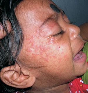
An 8-month-old boy presented with multiple facial lesions
present for the last 2 months, beginning as a cluster of
small papules on the right cheek. Examination revealed
multiple, 3-6 mm, erythematous, discrete as well as
confluent, firm papules distributed symmetrically over the
cheeks, chin, forehead, and eyelids (Fig. 1).
Routine hematological and biochemical tests, including lipid
profile were normal. Histopathology of a papule showed a
well-circumscribed collection of histiocytes in the upper
and mid-dermis. The cells were S100 and CD1a negative but
factor XIIIa positive. A diagnosis of benign cephalic
histiocytosis was made and the child was kept under periodic
follow-up.
 |
|
Fig. 1 Erythematous papules
of benign cephalic histiocytosis.
|
Benign cephalic histiocytosis is a rare,
self-limiting non-Langerhans cell histiocytic proliferative
disorder of young children, primarily affecting the face.
The average age of onset is 15 months. Asymptomatic
erythematous macules and papules on the face gradually
become reddish-brown and may spread to the neck, trunk, and
upper limbs; mucous membranes are not involved. The
differentials include Langerhans cell histiocytosis
(red-brown papules that show erosion, hemorrhage and
crusting), juvenile xanthogranuloma (orange/yellow papules
often with mucosal/ocular involvement), urticaria pigmentosa
(yellowish papules turning into weals on firm stroking), and
generalized eruptive histiocytoma (recurrent crops of
hundreds of yellowish/red-brown papules on face, trunk, and
extensors). The eruption clears spontaneously after a
variable period of time and requires no treatment.


