|
|
|
Indian Pediatr 2013;50: 512-513 |
 |
Primary Vertebral Lymphoma Presenting with
Fracture
|
|
Erman Atas, Vural Kesik, Erol Kismet and Vedat
Koseoglu
From Gulhane Military Medical Academy, School of
Medicine and Department of Pediatrics, Ankara, Turkey.
Correspondence to: Erman Atas, Gülhane Military
Medical Academy, Department of Pediatric Oncology, 06018 Etlik,
Ankara/Turkey. Email: [email protected]
Received: October 08, 2012;
Initial review: November 05, 2012;
Accepted: December 20, 2012.
|
|
We report a 15-year-old girl admitted with back pain and multifocal
osteolytic lesions without systemic symptoms at T7, L5, and S1 spinal
vertebras. The child was diagnosed as having primary multifocal osseous
lymphoma, in which multiple bones are involved in the absence of lymph
node or visceral disease for at least 6 months following initial
presentation.
Key words: Bone, Lymphoma, Vertebra.
|
Primary lymphoma of bone occurs rarely
in children and accounts nearly 2.8 to 5.9 percent of
Non-Hodgkin lymphomas [1,2]. The incidence of a single
vertebral lesion is reported to be 1.7% of all primary
lymphoma of bones [3]. Most of the involved bones are long
bones of the extremity, like femur [1]. The disease may
resemble fracture, trauma and mimic inflammatory,
neuropathic, and infectious conditions with these symptoms
[4,5].
Case Report
A 15-year-old girl was admitted with back
pain. On physical examination, there was tenderness on
thoraco-lumbar vertebraes. There was no history of trauma.
Lymphadenopathy, mass and organomegaly were not detected.
Laboratory data were as follows: Hb: 12g/dL, WBC: 6500/mm 3,
Platelet: 300000/mm3,
sedimentation: 14 mm / h, LDH: 146 U / L, renal and liver
function tests were normal. Thoracal vertebra X-ray
showed lytic lesions on T7 vertebrae. Thoracal computed
tomography (CT) showed reduced T7 vertebral corpus height,
and lytic, hypodense areas in the L5 and S1 vertebraes.
18F-Fluoro-deoxyglucose positron emission tomography
(18-F-FDG-PET) revealed increased activity on vertebral
corpus of T7, T11 and L4 vertebra and normal lungs. Bone
marrow aspiration and biopsy were normal. Pathologic
examination of the bone biopsy from T7 vertebra revealed
high-grade B-cell lymphoma. The patient was diagnosed as
primary bone lymphoma and LMB-89 chemotherapy treatment was
started. Three months later, magnetic resonance imaging
(MRI) showed heterogeneous hyper intense lesions on the
right side of sacrum, right iliac bone and acetabular roof
and left femoral neck, which were assessed as necrotic
lesions. 18-F-FDG-PET examination revealed increased FDG
uptake on right sacroiliac joint and sacrum; however, left
side had normal FDG uptake. Six months later, significant
improvement was detected on PET. The treatment was stopped 9
months later with no active lesion on bones. The patient is
now in remission for 66 months.
 |
|
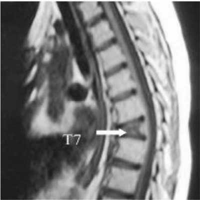
Fig. 1. T1 weighted
sagittal image of the thoracic spine shows wedge
shaped compression fracture of the thoracic 7
vertebra body. Spinal canal calibration is normal.
|
Discussion
Primary bone lymphoma is a rare disease
occurs primarily in the bone without an involvement of any
other site in the body. The most involved bones are femur,
tibia, mandible, mastoid, maxilla, zygomatic arch, rib,
clavicle, vertebrae, scapula, ulna, talus and calcaneous
[6,7].
The most common presenting complaints are
pain, swelling, mass, fever, weight loss, night-pain, limp,
irritability, pathologic fracture, and neurologic symptoms
[7]. The initial and only symptom in our case was back pain
due to vertebral fracture. Thus, in patients like our case
with limited symptoms; it is difficult to make differential
diagnosis. The mean delay from the onset of symptoms until
the final diagnosis was reported as 6.2 months (range, 0 to
2.5 years) [7]. The causes of delay were most often
nonspecific initial presentation like nonspecific pain
and/or swelling which can be attributed as musculoskeletal
pain, such as muscle strain or synovitis. Difficulty in
interpretation of the histological findings is a less
commonly reported reason of delay [7]. Our patient was
diagnosed 6 months after the pain began.
Pediatric primary bone lymphoma consists
of large cell lymphoma, lymphoblastic lymphoma, small,
noncleaved-cell lymphoma, and unclassified [6]. Pediatric
diffuse large cell lymphoma has a favorable prognosis from
others [8]. Back pain and vertebral fracture are the two
complaints that can be commonly seen in children with
trauma, arthritis, and infections. This can lead serious
delay in diagnose. Unresolved pain and fracture despite
analgesic treatment may be a good pointer to the possibility
of a lymphoma.
Contributors: All the authors have
contributed, designed and approved the study.
Funding: None; Competing
interests: None stated.
References
1. Furman WL, Fitch S, Hustu HO, Callihan
T, Murphy SB. Primary lymphoma of bone in children. J Clin
Oncol. 1989;7:1275-80.
2. Anderson JR, Wilson JF, Jenkin DT,
Meadrows AT, Kersey J, Chikote RR, et al. Childhood
non- Hodgkin’s lymphoma: the results of a randomized
therapeutic trial comparing a 4-drug regimen (COMP) with a
10-drug regimen (LSA2-L2). N Engl J Med. 1983;308:559-65.
3. Huang B, Li CQ, Liu T, Zhou Y. Primary
non-Hodgkin’s lymphoma of the lumbar vertebrae mimicking
tuberculous spondylitis: a case report. Arch Orthop Trauma
Surg. 2009;129:1621-5.
4. Bhagavathi S, Fu K. Primary bone
lymphoma. Arch Pathol Lab Med. 2009;133:1868-71.
5. White LM, Siegel S, Shin SS, Weisman
MH, Sartoris DJ. Primary lymphoma of the calcaneus. Skeletal
Radiol. 1996;25:775-8.
6. Suryanarayan K, Shuster JJ, Donaldson
SS, Hutchison RE, Murphy SB, Link MP. Treatment of localized
primary non-Hodgkin’s lymphoma of bone in children: a
Pediatric Oncology Group study. J Clin Oncol. 1999;17:456-9.
7. Glotzbecker MP, Kersun LS, Choi JK,
Wills BO, Schaffer AA, John P. Dormans primary non-hodgkin’s
lymphoma of bone in children. J Bone Joint Surg Am.
2006;88:583-94.
8. Zhao XF, Young KH, Frank D, Goradia A, Glotzbecker MP,
Pan W, et al. Pediatric primary bone lymphoma-diffuse
large B-cell lymphoma: morphologic and immunohistochemical
characteristics of 10 cases. J Clin Oncol. 1989;7:1275-80.
|
|
|
 |
|
