|
|
Case Reports Indian Pediatrics 2005; 42:479-482 |
||||
|
Plasmapheresis in Childhood Acute Disseminated Encephalomyelitis |
||||
|
Rajesh RamachandranNair From Department of Neurology, Medical College Hospital, Kozhikode, India. Correspondence to: Rajesh RamachandranNair, Division
of Neurology, The Hospital for Sick Children, University of Toronto, 555
University Avenue, Toronto, Canada M5G 1X8. Manuscript received: June 29, 2004; Initial review completed: November 1, 2004; Revision accepted: December 13, 2004.
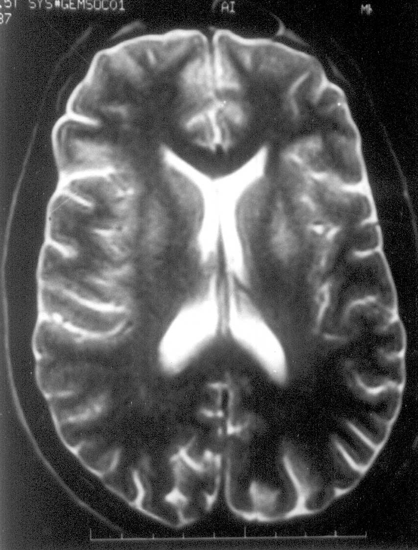
Acute disseminated encephalomyelitis (ADEM) is an acute inflammatory de-myelinating disease of the central nervous system, typically characterized by onset of multifocal neurological deficits days to weeks after an episode of viral illness or vaccina-tion(1). Accepted methods of treatment include intravenous methylprednisolone (IVMP), intravenous immunoglobulin (IVIG) or a combination of both(2-4). There are a few reports highlighting the importance of plasmapheresis in ADEM(5,6). We report two cases of ADEM in children treated effectively with plasmapheresis. Case Reports Case 1: A 12-year- old girl developed right focal seizure 10 days following an upper respiratory tract infection. On the second day she developed progressive difficulty in walking. She had mild dysarthria and dysphagia. She also complained of headache. On the fifth day, she became completely bed-bound with altered sensorium. The Glasgow Coma Score (GCS) was 11 (E3 V4 M4). Neurological examination revealed a normal retinal fundus and bilateral pyramidal signs with quadriplegia. There was mild bilateral upper motor neuron type facial palsy and the palatal movements were decreased. Hemogram, renal and liver functions, chest X-ray and electrocardiogram were normal. Blood and urine cultures did not grow any organism. The following tests were negative or normal: CRP, ANA, Widal test, peripheral smear for Malarial parasite, serum HIV-ELISA, serum antibody test for Ebstein Barr, measles, mumps, rubella, herpes simplex and Japanese encephalitis viruses, Brucella and Myco-plasma. Electroencephalogram showed diffuse slowing without epileptiform dis-charges. MRI brain on the 6th day of illness showed multiple hyperintense lesions in the sub cortical white matter, right basal ganglia and internal capsule in T2 weighted images, suggestive of demyelination (Fig. 1). CSF showed white cell count of 76 per cumm(90% lymphocytes), 64mg% protein and 80 mg% glucose. CSF PCR for Herpes simplex virus and antibody for Japanese encephalitis virus were negative. She was treated with IVMP at a dose 30 mg/kg body weight per day for five days followed by oral prednisolone at a dose of 1mg/kg body weight for two weeks. There was no significant improvement following steroid therapy. On the 25th day of illness, she was still bed bound with a GCS of 9. Parents could not afford IVIG. Hence plasmapheresis was initiated on 26th day of illness. A total six exchanges were done over a period of two weeks removing 240 mL/kg body weight plasma. Fresh frozen plasma and isotonic saline were used as replacement fluids. Clinical improvement began from the third exchange onwards. At the end of six exchanges she had a remarkable recovery. The only neurological deficit at the end of plasmapheresis was mild dysarthria with hyperreflexia. But she could walk without support. One month after the procedure she had no neurological deficit. Repeat MRI brain at 3 months showed total clearance of the brain lesions (Fig. 2).
Case 2: A 14-old-boy was admitted to the neurology ward with ataxia, dysarthria and weakness of left side. Symptoms began 4 days following a short febrile illness. He was admitted on the 3rd day of illness. He did not have seizures or alteration in the level of consciousness. Examination showed bilateral pyramidal and cerebellar signs with sluggish pharyngeal movements. He could not walk even with support. Routine blood examination was normal. Computerized tomogram of the brain (without contrast) on the third day did not reveal any structural lesions. CSF examination showed, a total leucocyte count of 46 cells per cumm with 86% lymphocytes and 14% polymorphs, protein 61mg% and glucose 73 mg%. Electroencephalogram was normal. MRI brain (5th day of illness) showed multiple hyperintense lesions in T2 W images involving subcortical white matter, right thalamus, basal ganglia and internal capsule. He was treated with IVMP at a dose of 1 g per day for five days, followed by oral prednisolone (1mg/kg body wt) for two weeks. On the 23rd day of illness he was bed bound due to ataxia and weakness. As the parents could not afford IVIG plasmapheresis was started on the 24th day of illness. A total of five exchanges were done over a period of 10 days. 50 mL/kg body weight of plasma was removed in each sitting. Fresh frozen plasma and isotonic saline were used for replacement. From the second exchange onwards there was improvement in the neurological deficits. At the end of 5 exchanges he had only mild left hemiparesis and subtle ataxia. One month after the procedure, he had a normal neurological status. Repeat MRI brain at 3 months showed clearance of the lesions. Discussion Diagnosis of ADEM was made on the basis of clinical picture, CSF findings and the MRI features. These two children had no significant clinical improvement after steroid therapy. In steroid resistant ADEM, the currently used therapies include plasma-pheresis and IVIG(2-4). Efficacy of one over the other is not yet established. The practice followed in children is to try plasmapheresis, if the response to steroid and IVIG was not satisfactory. This is probably because of the ease of administering IVIG with less risk. Since parents could not afford IVIG, we planned plasmapheresis. Plasmapheresis was used after a course of steroid in all the series and case reports(5-7). Thus a delayed effect of steroid cannot be excluded. Experimental autoimmune encephalomyelitis, induced by inoculating animals with myelin or myelin products is a good model for ADEM(8). If ADEM is considered as a cell mediated immune mediated disease it would be difficult to explain the use of plasmapheresis in ADEM. It is believed that the effectiveness of plasmapheresis is due to its ability to remove offending circulating antibodies(9). There is no consensus regarding the total volume of plasma to be removed in ADEM. Tselis(2) suggested a total of six or seven exchanges every 2 days, each plasma exchange consisting of removal of approximately one-plasma volume. Miyazawa, et al.(6) treated an 11-year-old with 4 sessions of plasmapheresis (average 54 ml/kg). We used a similar dose and the effect was significant and sustained. There could be individual variation in the frequency, duration and volume of plasma exchange. There is report of a lady who relapsed twice after the end of plasmapheresis, but had responded to repeat plasmapheresis(9). These two cases demonstrated that plasmapheresis could be useful even in the fourth week in severe ADEM not responding to steroid therapy. Plasmapheresis is an option for IVIG resistant cases and those who cannot afford IVIG. A randomized control trial is needed to address issues like the volume, frequency, duration and timing of plasmapheresis in ADEM. Contributors: RR was in charge of patient care, supervised plasmapheresis and prepared the manuscript. MR did the plasmapheresis and collected the literature. ASG edited the manuscript. Funding: None. Competing interests: None stated.
| ||||
|
References | ||||
|
|
![]()