|
|
Letters to the Editor Indian Pediatrics 2000;37: 801-802 |
|
|
Pitfalls in the Diagnosis of Esophageal Atresia |
|
|
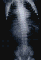
A term male neonate weighing 2.1 kg weight presented on the 4th day of life with drooling of saliva and regurgitation of feeds since birth. The infant was tachypneic and had chest retractions. Esophageal atresia was suspected and an 8Fr nasogastric tube was passed. This went down into the stomach as confirmed on a plain X-ray (Fig. 1 ). The child was treated for pneumonitis and sepsis. Because of the persistence of excessive oral secretions, reassessment of the infant was done. A non-ionic contrast study was performed which confirmed the presence of esophageal atresia. Primary repair was carried out. The infant developed an anastomotic leak on the third post-operative day. This was followed by respiratory failure and sepsis to which he succumbed after 35 days of intensive care. The delay of 24 hours in diagnosis occurred because the catheter had passed into the stomach inspite of the esophageal atresia. The soft catheter probably went through the trachea and from there into the stomach through the fistula. Esophageal atresia is one of the common major congenital emergencies and needs a prompt confirmation of the diagnosis. The simplest and safest way to settle the question is to pass a stiff radio-opaque catehter down the mouth and into the stomach and confirm the position by chest X-ray(2). If a stiff tube has reached the stomach, it can be safely assumed that there is no esophageal atresia. Also. It is unlikely to pass accidentally through the trachea into the stomach via the fistula. In order to prevent this mistake, stiff (10 Fr) catheter should be used for the diagnosis of esophageal atresia. Alka Gupta,
|
![]()