|
|
Images in Clinical Practice Indian Pediatrics 2005; 42:390 |
||
|
Solitary Mastocytoma |
||
|
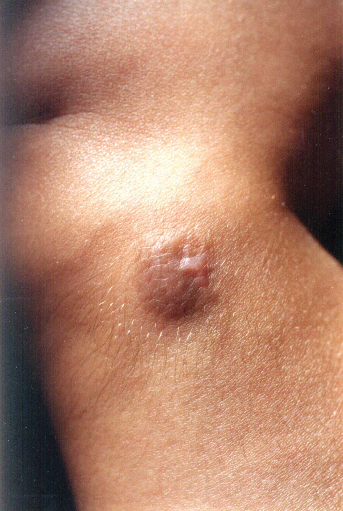
Mastocytoma usually appear as solitary lesion, although it is estimated that 10% to 15% of all patients with one of the mast cell disorders (urticaria pigmentosa, telangiectasia macularis eruptive perstans, diffuse mastocytosis, systemic mastocytosis, and mast cell leukemia) have a mastocytoma. They appear as pink-yellow to tan, round to oval nodules or plaques, 0.5 cm to 3 cm in largest diameter. Mastocytomas have a smooth or peau d’orange surface with a rubbery consistency. The common locations of mastocytomas are on the trunk, neck, and arms. The lesions appear at birth or in the first few months of life and rarely occur later in life. Mastocytomas may precede another form of cutaneous mast cell disease or be associated with attacks of generalized flushing and abdominal colic. The diagnosis of mastocytomas is less easy, the clinical differential diagnosis includes melanocytic naevi, xanthomas, or juvenile xanthogranulomas. A positive Darier’s sign which may be observed in only half the cases, suggests the diagnosis of mastocytoma. The course of solitary mastocytomas is benign. Those lesions not cured by excision appear to improve or resolve during early childhood. Symptomatic therapy of cutaneous mastocytosis involves agents that inhibit the release of mediators or antagonize H1 and H2 receptors such as antihistamines, ketotifen and aspirin. Skin-targeted therapies that lead to a resolution of the lesions of cutaneous mastocytosis are psoralen-photochemotherapy and topical corticosteroid therapy either by occlusion or intralesional injection for a limited number of lesions. Devinder Mohan Thappa, |
![]()