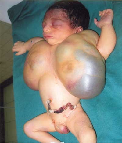
A term newborn presented with large, well defined swellings, approximately
8cms × 10cms, bilaterally over the anterior chest wall. Overlying skin
showed bluish discoloration. Multiple small bluish swellings were also
present on the right upper limb (Fig 1). A diagnosis
of multiple hemangiomas with giant chest wall hemangiomas was made. No
visceral lesion was found on ultrasonography. Baby died on third day of
life due to haemorrhage and shock. Biopsy findings were consistent with
hemangioma.
 |
|
Fig.1 Giant bilateral chest wall
hemangiomas. |
Hemangiomas occur in 1.1 to 2.6% of the neonates . They
can be classified as superficial , deep or mixed. Deep hemangiomas need to
be differentiated from lymphatic or venous vascular malformations. MRI can
help distinguish between a vascular malformation and hemangioma.
Majority of hemangiomas involute without intervention.
Giant hemangiomas can however present with complications such as cardiac
failure, haemorrhage, platelet trapping and disseminated intravascular
coagulation and may require early intervention. Modes of therapy employed
include surgical excision, steroids, embolisation, radiotherapy and
pneumatic compression. An early surgical intervention may have improved
the outcome in this neonate.


