|
|
|
Indian Pediatr 2020;57:
983 |
 |
Targetoid Hemosiderotic Hemangioma
|
|
Avik Panigrahi* and Abheek Sil
Department of Dermatology, Venereology, and Leprosy, RG Kar
Medical College, Kolkata, West Bengal, India.
Email: [email protected]
|
|
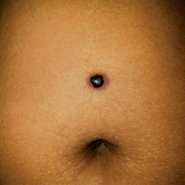
A 10-year-old boy presented with 1-year history of a
gradually progressive non-tender, soft-to-firm, dome-shaped,
brownish-black papule (6x6 mm) with a peripheral erythematous halo
situated above the umbilicus (Fig. 1). There was no
history of preceding trauma, acute illness or any drug intake. Other
mucocutaneous areas were uninvolved. Excision biopsy confirmed the
clinical impression of targetoid hemosiderotic hemagioma (THH); no
recurrence was noted on regular follow-up.
 |
|
Fig. 1 Targetoid hemosiderotic
hemagioma characterized by a dome-shaped, brownish-black papule
with surrounding erythe-matous halo.
|
THH is an acquired benign vascular lesion presenting
as a solitary, red-violaceous to brown targetoid papule with a
hemorrhagic halo; usually adolescent onset. Classic histology shows
biphasic pattern: dilated vessels lined by hobnail endothelial cells
with intraluminal papillae in the papillary dermis; and angulated and
slit-like vascular spaces dissecting the collagen bundles in the
reticular dermis, with plenty of extravasated erythrocytes and
hemosiderin deposition at the periphery (accounting for the targetoid
appearance). They are often misdiagnosed as melanocytic nevus (coarse
hair, absence of halo, presence of melanocytic nests), infantile
hemangioma (bright red lobulated plaque with typical growth pattern),
dermatofibroma (painful, positive dimpling sign), solitary angiokeratoma
(no halo, hyperkeratosis and dilated vessels only in papillary dermis on
histology) or melanoma (rare in pre-pubertal age, atypical melanocytic
nests). Complete removal is sufficient to treat the condition.
|
|
|
 |
|
