An 8-month-old female with a history of eye discharge,
presented with complaints of pustules on red tender skin,
which ruptured to lead to erosions and peeling since 3 days.
On examination, skin was tender with diffuse erythema.
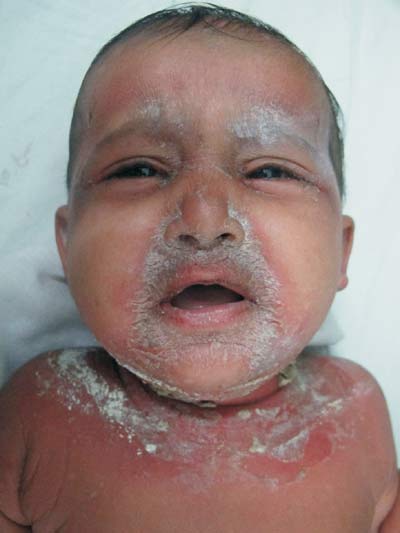
Whitish crusting and fissuring was seen in the perioral area
and the neck (Fig. 1), with sparing of the mucosa.
Flaccid pustules and blisters, few having ruptured to lead
to erosions were seen on trunk, inner thighs and neck.
Nikolsky sign was positive. Wrinkling of skin along with
exfoliation was seen in the axillae. There was leucocytosis.
Lesional pus for smear and culture sensitivity and blood
culture were negative for Staphylococcus.
Histopathology revealed focal loss of upper epidermis and
presence of acantholytic cells in the subcorneal layer. A
diagnosis of staphylococcal scalded skin syndrome (SSSS) was
made. Patient had a complete recovery with peeling within 10
days on treatment with antistaphylococcal antibiotics.
 |
|
Fig.1 Whitish crusting and
fissuring in the perioral and perinasal areas with
flaccid blisters in neck folds.
|
SSSS, caused by Staphylococcus aureus
exfoliative toxins (ET) A and B, generally affects neonates,
infants, and children less than 5 years of age, due to lack
of protective antitoxin antibodies and immature renal
function. Left untreated, large sheets of epidermis slough
off to leave extensive areas of raw denuded skin that is
sensitive and painful. The toxin is usually produced at a
site distant from the lesions. ET acts as an atypical
glutamate-specific serine protease that binds and cleaves
desmoglein-1(found in the upper epidermis, absent in the
mucosa) which explains the specific site of action in the
superficial epidermis and the absence of mucous membranes
affection in SSSS. Cultures from the skin lesions are
negative for staphylococcus in almost all cases. It is
important to send swabs from other areas such as the
umbilicus, nasopharynx and conjunctivae. Anti-staphylococcal
antibiotics, temperature regulation, maintaining fluid and
electrolyte balance, nutritional management and skin care
form the basis of treatment. The main differential diagnosis
remains drug-induced toxic epidermal necrolysis (TEN) the
differentiating factors in TEN being-adult onset, spared
areas of the skin, mucosal involvement, presense of nikolsky
sign only in involved skin (and not diffusely) and absence
of perioral/perinasal crusting.
It is important to recognize this often
dramatic looking skin disorder early, especially in
nurseries, with the help of the above-mentioned classical
features.


