|
|
Personal Practice Indian Pediatrics 2002; 39:922-930 |
||
|
Atopic Dermatitis |
||
|
Atopic dermatitis (AD) or eczema is a common chronic or relapsing dermatitis characterized by severe pruritus, occurring primarily in infants and children(1). It affects 5-15% of school children and 2-10% of adults(2,3). Atopic dermatitis is notorious for its recalcitrance and tendency to chronic recurrence and can lead to significant morbidity, social isolation and emotional stress. Epidemiology There is increasing evidence that the prevalence of atopic dermatitis in children has increased over the past 30 years(4,5), although the reasons for this increase are unknown. The current prevalence is estimated to be between 10.0% to 15.6%(6). Changes in environmental pollutants, breastfeeding pattern, increased awareness, urbanization and better case detection techniques are some of the reasons cited for this change(4,7). A similar trend has been observed in India over the past 30 years(8). In an Indian study from Bihar in 1972(9), the overall incidence of AD was 0.38% of the total number of cases of skin diseases; of these, 38% had "infantile AD". In another study done two decades later, AD comprised 28.46% of the total pediatric skin diseases(10). The disease starts early with 35% to 60% of symptoms manifesting in the first year of life and 47% to 85% by 5 years of age(11). Most of the Indian children develop the disease in infancy(10). Although females outnumber males with a ratio of 3:2(12), male preponderance has also been observed both in India and the west (10,13). The disease is known to exacerbate in winter and improve in summer months(12). A direct relationship has been observed between socio-economic class and the prevalence of AD in children(14). Etiopathogenesis The disease arises as a result of a complex interplay between various genetic, immunological and environmental factors (5,13). Atopic dermatitis clearly has a hereditary basis(4,5,15). The eczema is triggered or exacerbated by interactions between a genetic predisposition and environ-mental factors(4,15). The environmental factors include (a) physical factors like sweating, climate, warm surroundings, detergents and soap, synthetic or woollen fabrics, cigarette smoke, (b) psychological factors, (c) food items (including tomato, orange and citrus fruits, juice from meat, fish) (d) allergens such as house dust mite, animal hair, pollen, plants and others such as Staphylococcus aureus and release of exotoxins (superantigens) and saliva in small children(15). Majority of cases are associated with a sensitization to environmental allergens and increased serum IgE (extrinsic AD), but about 10-30% of all cases lack any link to the classical atopic diathesis and are labelled as intrinsic AD(5,16).
Diagnosis There is no laboratory gold standard for the diagnosis of AD. A detailed history and a characteristic clinical picture would establish the diagnosis. Hanifin and Rajka(17) have laid down certain major and minor criteria for making a diagnosis of AD (Table I). However, both Western and Indian Studies have shown that the various minor clinical features of AD vary in specificity in different age groups of the patients(18,19). Thus, the UK Working Party(20) modified these criteria and included ‘xerosis’ as well as ‘early age of onset’ as minor features. Incidentally, these two were the most significant features found in Indian studies(19). In case, a child presents with some of the minor clinical features without the presence of eczema, the condition is labelled as "atopic diathesis" and such patients have a strong tendency to develop AD in future(8). Clinical features Pruritus is considered to
be the primary phenomenon. However, AD can also begin in the first three
months of life before the appearance of co-ordinated scratching(21).
Xerosis, or dryness of the skin can be found shortly after birth and can
act as the inciting factor for AD. Lesions are classified as acute,
subacute or chronic. Acute AD is characterized by pruritic papules and
papulovesicles with serous exudates on a background of erythema.
Subacute eczema is characterized by either grouped or scattered scaly,
erythematous papules or plaques over an erythematous skin. Chronic AD
includes thickening of the skin with lichenification (increased skin
markings), secondary to scratching and rubbing. Usually in dark skinned
individuals, the lesions may sometimes have a perifollicular
distribution, giving the skin a pebbled appearance or the seemingly
unaffected skin may have a dry lacklustre appearance(9).
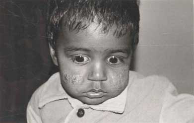
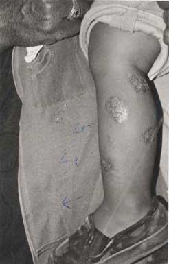
The distribution of lesions varies with age(1). Face and scalp involvement is common in infants (Fig.1) as well as the extensor surfaces of the extremities and the trunk. Infants with extensive facial involvement may rub their face on the sheets of the crib or the shoulder of the person holding them(4). Cradle cap has also been observed on the scalp in a large Indian series(10). This is known as the infantile phase of AD. In the childhood phase of AD 18-24 months onwards, eczema is observed on the flexural surfaces, including the neck, antecubital or popliteal fossae, wrists and ankles. Acute, generalized or localized vesiculation should always suggest the possibility of secondary bacterial or viral infection (Fig. 2).
There is an adult phase, which occurs in adolescents and adults, where lichenification of the flexures and the hands commonly occurs. Hand involvement is a common feature in patients with AD varying from 10 to 70% in different series(10,21). Some of the commonly observed minor features of AD are: Xerosis: Dry looking skin with fine scaling and perifollicular accentuation(1,4). Ichthyosis vulgaris: Characterized by polygonal fish-like scales on the extensor surfaces. Keratosis Pilaris: Asymptomatic keratotic follicular papules on the extensor surface of the upper arms, buttocks, and anterolateral thighs. Pityriasis alba: Hypopigmented scaly patches on the cheeks and rarely on the upper arms and shoulders(4). Dennie-Morgan fold: Single or double fold in the lower eyelid. Atopic dermatitis may be complicated by bacterial infections (staphylococcal), viral (herpes simplex), or superficial fungal infections. Other complications include erythroderma and psychomotor symptoms because of social ostracization. Several factors seem to influence the severity of AD including racial and ethnic considerations, country of origin, environmental factors, emotional factors, breast feeding, weaning and dietary habits(4,22). The severity can be measured using Rajka and Langeland’s scoring system(23). Atopic dermatitis in India is relatively milder than in the west(10,22). The reason for this has been attributed to warmer climatic conditions, dietary habits and clothing materials used, prolonged breast feeding, the low frequency of personal and family history of atopy and the low incidence of colonization of atopic skin by Staphylococcus aureus(22). Management The treatment is aimed at suppressing the symptoms and controlling or preventing complications (Table II). First line treatment Explanation and discussion: An important aspect in the management of the disease is to spend adequate time with the parents of the atopic child explaining the nature of the disease, and what to expect realistically from the treatment. Explanation and education regarding the application of topical preparations and the quantity to use (Table III), avoidance of exacerbating factors and caring for the sensitive skin of the child including bathing and emollients should be given(3). Avoidance of provoking factors: Soaps and detergents remove the natural lipids from the surface of the skin and hence their use should be minimized(3,4). Extremes of temperature should be avoided and woollens should not be worn next to the skin(15). Cotton clothing is to be preferred. Stress should be avoided(1,4). Bathing and emollients: Bathing and moisturizing is most useful for cleansing and hydrating the skin(4) .For bathing, only mild soaps without any fragrance or a non-soap cleanser (e.g. Cetaphil) or a soap containing a moisturiser should be used. This should be immediately followed by patting the body dry with a towel and applying emollients(4,24). Emollients such as vegetable oils or petrolatum jelly provide a surface lipid film which retard evaporative water loss from the epidermis. Emollients and topical medication should be applied within 3 minutes after bath to retain hydration (15). Emollients should be reapplied to hands and face at regular intervals during the day. Antibiotics: Antibiotics should be used for treating overt secondary bacterial infection. In an Indian study, oral erythromycin and cloxacillin given for 21 days in children with atopic dermatitis led to a significant improvement in eczema and pruritus along with the drop in the CFU/cm2 of Staphylococcus aureus(25). Prophylactic treatment of staphylococcal carrier sites (such as the nose, axillae and perineum) with topical antibiotics such as mupirocin may be appropriate in patients with recurrent infected eczema(3). Topical corticosteroids: Besides emollients, topical corticosteroids are the mainstay of treatment for AD and can be used safely if certain precautions are taken (Table III). The choice of different potencies of topical corticosteroids will depend on the status, site and extent of eczema(3). The risks and benefits of the treatment should be explained to the parents. One percent hydrocortisone ointment is adequate for most infants with mild to moderate eczema and should be used on the face. New topical steroids which can be effective even when applied once a day are relatively safe e.g., desonide, prednicarbate, fluticasone propionate and mometasone furoate(26). A high potency corticosteroid can be used for a short period to control acute flares of the disease(15). Dry, lichenified AD may require a mid potency steroid for 2-3 weeks such as betamethasone dipropionate ointment 0.025% twice a day. If there is superadded bacterial or fungal infection, a topical steroid/antibiotic or steroid/antifungal preparation should be used. Adverse effects of topical corticosteroids include atrophy, depigmentation, steroid acne, telangiactesia and rarely suppression of the hypothalamic-pituitary-adrenal axis(15). There is also an increased risk of contact irritant dermatitis due to application of topical steroids and medicaments containing their combination with antibiotics and antifungals, due to ingredients used as preservatives which act as sensitizers e.g., paraben, benzyl alcohol, chlorocrenol, isopropyl palmitate, polysorbate 60, stearyl alcohol, neomycin, fragrance, local anesthetics(27). It is recommended that such medications should be used carefully. Compresses: Wet compresses of plain tepid water or normal saline when applied on oozing skin remove debris and cool inflamed skin and are useful for severe, exudative lesions. Wet dressings: These are especially useful in infants and children with severe atopic dermatitis(1). A wet dressing is applied directly to the skin and left in place for several hours following application of a cream or an ointment(1). There are many modifications of this general approach which further increases penetration of the topical medication (4) but usually a double layer of either gauzes, wraps, towels or stockinettes or cotton pajamas are placed on the patient over the affected areas, the inner layer being wet (moistened with warm water) and the second being dry (1,3,4). This method can provide good symptomatic relief. Antihistamines: They are used for their sedating properties to enhance sleep and to made pruritus more tolerable(15). Sedative antihistamines such as hydroxyzine and promethazine are preferred to non-sedative ones. They should be used at night, an hour or so before bedtime in all those children who have difficulty in sleeping, or scratch while asleep. Psychological support: A psychologist may be able to help the child and the parents by detailed discussion, behaviour therapy or relaxation therapy (9,15). Second Line treatment Avoidance of allergens: Regular cleaning and changing of mattresses, keeping simple furniture, avoidance of carpeting at home and regular washing of old, soft, furry toys can help the child with severe AD(24). A trial of dietary manipulation is to be strictly avoided in a developing country like India where malnutrition is a major health concern, unless the patient’s history is strongly suggestive of a specific food allergy(8). Before omitting a food item in suspected cases of food allergy, it is necessary to carry out a RAST combined with double blind placebo controlled oral food challenge(8). Breast feeding does seem to have a protective effect in relation to severity during the early months of life and should therefore be encouraged. Systemic corticosteroids: Systemic corticosteroids have a limited role in tiding over occasional flares of severe AD but the decision to use them should be taken after serious consideration as they may have detrimental effects on the growth of a child or an adolescent. Other treatment modalities: Phototherapy and photochemotherapy, cyclosporin, evening primrose oil, azathioprine, Chinese herbal medicines, interferons, thymopentin, human interferon gamma and plasmapheresis have been found to be effective in AD, but they are used mainly in the west. As we see a relatively milder form of the disease in India, these modalities may not be required for our patients. In our pediatric dermatology clinic, we manage the patients by advising them to avoid provoking factors, to keep nails short to apply topical mupirocin for carrier sites, systemic antibiotics for infection ,topical corticosteroids and for severe flares, systemic corticosteroids for short periods. There is limited experience with photochemotherapy, phototherapy and cyclosporine administration. Recent Advances In infants at high risk of developing atopic disease, breastfeeding in conjunction with maternal hypoallergenic diet appear to decrease the prevalence of AD(28), however the benefits may not be sustained. Scientific evidence to support the use of homeopathic remedies in atopic eczema is still awaited. Topical FK506 ointment or tacrolimus, a member of the macrolide family of immunosuppressive medications, such as cyclosporine has been shown to be effective in the treatment of AD in clinical tria1s(29). Its mechanism of action seems to be mediated through binding to cytosolic binding proteins, known as immunophilins, binding to antigen-presenting epidermal cells, such as langerhans cells and down regulating the expression of the high-affinity receptor for IgE(29). It is not as yet available in India. Topical SDS ASM 981, a macrolactam ascomycin, which is an inflammatory cytokine inhibitor also appears to be a promising prospect(30). Table II_ Management of Atopic Dermatitis First Line Treatment • Explanation and discussion with parents • Avoidance of provoking factors • Bathing and emollients • Treatment of infection with antibiotics • Topical corticosteroids • Tar preparations • Antihistamines • Psychological support Second Line Treatment • Allergen avoidance • Systemic corticosteroids
• Phototherapy or photochemotherapy Other Therapies • Tacrolimus (FK 506) • Ascomycin • Essential fatty acid supplements • Psychotherapy • Traditional Chinese herbal medicines • CD4 monoclonal antibody • Photopheresis Table III_ Topical Steroids in the Management of Atopic Dermatitis • Age: For infants and children with mild to moderate eczema and for sites such as the face, mild potency steroid such as I % hydrocortisone ointment is adequate. In older children, a moderately potent steroid such as betamethasone dipropionate, 0.025% cream can be used. • Sites: As corticosteroid penetration is increased on face and flexures, mild steroid preparations should be used. Even in the eyelid area, potent steroids should be avoided to prevent the development of long term complication of glaucoma. • Extent of eczema: The potential for systemic absorption increases with the extent and activity of the eczema which could lead to risk of suppression of hypothalamic-pituitary -adrenal axis, so monitoring of use of steroids in severe AD is important. • Type of preparation: Ointment bases are preferable to cream bases as they provide more occlusion and penetration but in humid environments, may lead to folliculitis as they are more greasy. Thus creams may be better tolerated in some instances. • Method of application: Topical steroids should not be applied more than twice daily and some of the newer preparations only once a day. Cover the eczematous areas evenly with a fine film of ointment so that the surface of the skin glistens in the light. The amount to apply can be explained to the parents in terms of fingertip units, where one fingertip unit equals 0.5 g .The amount of potent topical steroid applied on a small child should not exceed 10-20 g. weekly to avoid hypothalamic-pituitary-adrenal axis suppression. Course Atopic dermatitis follows a highly variable course with exacerbations and remissions. About 95% of children with AD remit around puberty, but relapses may occur and the disease may persist well into adulthood. With severe AD, in 72% the disease persists in adult life(31 ). Risk factors regarded to affect the disease prognosis include severe dermatitis in childhood, family history of AD, associated asthma or allergic rhinitis, female sex and onset before 1 year of age(32). Contributors: RS reviewed the literature and drafted the paper. AJK provided the concept and critically revised the manuscript. RS shall be the guarantor for the paper.
| ||
|
References | ||
|
![]()