|
|
|
Indian Pediatr 2013;50:
1020-1024 |
 |
Birthweight Centile Charts from Rural
Community-based Data from Southern India
|
|
Anu Mary Alexander, Kuryan George, Jayaprakash Muliyil, Anuradha Bose
and Jasmin Helan Prasad
From Department of Community Medicine, Christian Medical College,
Vellore, Tamil Nadu, India.
Correspondence to: Dr Anu Mary Alexander, Department of Community
Medicine, Christian Medical College, Vellore, 632 002, Tamil Nadu,
India.
Email:
[email protected]
Received: December 14, 2012;
Initial Review: February 5, 2013;
Accepted: May 7, 2013.
Published online: June 5, 2013.
PII: S097475591201057
|
Objective: The objectives of the study were to estimate gestational
age specific birthweight centiles from healthy pregnancies in a defined
rural block and compare the under-two month mortality rates in those
belonging to the lowest and highest centile groups.
Design: Retrospective chart review.
Setting: Routine data collected regarding all
pregnancies, births and deaths occurring in Kaniyambadi, a rural block
in Southern India, between 2003 to 2012.
Subjects: All singleton live newborns of women
without known major antenatal risk factors.
Main outcome measures: Gestational age- and
sex-specific birthweight centile curves were created using the LMS
method. Mortality rates for the first two months of life were calculated
for those in various centile groups.
Results: The median birthweight at term was lower
for the study subjects as compared to the median birth weights in the
WHO child growth standards 2006, the US and the UK standards. Mortality
rates for those with birthweights both below the 3rd centile as well as
above the 97th centile higher than for those between 3rd and 97th
centiles.
Conclusions: While absolute values of
birthweights were lower than the WHO 2006 child growth standards there
was a J shaped curve of birthweight and mortality. This suggests that in
a given population, mortality increases at extremes of birthweights,
even if some of these birthweights may be considered normal by other
standards.
Keywords: Birthweight, India, Mortality, Outcome, Rural.
|
|
Birth weight centiles are available for developed
countries [1,2] but are not easily available in developing countries
although the importance of ethnic-specific standards has been
acknowledged [3]. Sex- and parity-specific birthweight charts for
gestational age have been published based on births in tertiary
hospitals but community-based birth weight centile charts representing
all births in a population are not available [4,5]. An earlier
comparison of birth weights in rural south India against an Indian
standard showed that the Indian standard was descriptive rather than
normative as the proportion small-for-gestation (SGA, below the 10th
percentile) was much lower than the proportion who were low birth weight
(below 2500g), while comparison with a Canadian standard showed a high
rate of SGA [6].
The primary objective of this study was to construct
birthweight centiles for children born to healthy mothers in a rural
block in southern India between 2003-2012 and to compare with the birth
weights of developed countries as well as the WHO child growth
standards. The secondary objective was to compare mortality rates within
two months of birth, for children with birth weights below the 3rd
centile and above the 97th centile to those with birthweights between
the 3rd and 97th centiles.
Methods
The study was carried out in a rural block of 110 000
population and 82 villages in southern India, which is the service area
of the Community Health and Development (CHAD) program linked to the
community health department of a large tertiary hospital. The primary
care activities of the program are supported by a 140-bedded secondary
hospital. Information regarding pregnancies, births and deaths in this
area is collected and maintained in a computerised database by female
health workers as described in an earlier study [6].
The prevalence of low birth weight (below 2500 g)
and preterm births (<37 weeks) in this area was 17% and 5.5%,
respectively while proportion of women receiving any antenatal care was
99% in 2005 [6].
The data on births occurring in the block was
obtained from the computerised database maintained by the health
information system of the secondary hospital [6]. Only births with
recorded birth weights and gestational age at the time of delivery
(determined by either the date of the last menstrual period or
ultrasound scans) were included in the final database. Tukey’s method
was followed to exclude extreme values/outliers of birthweights, which
revealed seven outliers [7]. The upper limit (Tukey’s outer fence) was
taken as the third quartile value plus 3 times the interquartile range
while the lower limit of normal (Tukey’s inner fence) was taken as the
first quartile minus three times the interquartile range.
As the birthweight centiles were meant to be
descriptive of healthy pregnancies, mothers diagnosed with medical
conditions known to have an effect on birthweight, including gestational
diabetes, pregnancy induced hypertension, maternal heart disease, short
stature (taken as maternal height less than 140 cm), and hemoglobin less
than 10 g/dL were excluded [8].
The data were used to compute smoothed centile
charts, based on the LMS method [9,10] using the software LMS Chartmaker,
according to sex of the child and parity of the mother. This method uses
the Box-Cox Power transformation to make the distribution normally
distributed and produces curves of L (Box-Cox power parameter), M
(median) and S (coefficient of variation) plotted against age. The
centiles drawn were 3 rd, 10th,
25th, 50th,
75th, 90th
and 97th and spread sheets
with L, M, S values and centile values were also created. For each
child, the Z score was computed using the previously described formula
[11].
Although most infant deaths occur in the first month
of life, we used under-two month mortality rates as a means of
validation of the birth weight centile groupings, in order to account
for all deaths may be associated with low birth weight. The mortality
rates were computed using available mortality data for children born to
permanent residents of the study area, as follow up was done mainly for
these children and not for mothers who were temporary residents (mothers
who had only come for antenatal care/delivery to their maternal homes).
The number of deaths in various centile groups among the children
included in the birth weight centile data were used for obtaining these
rates, thus giving under two month mortality rates for singleton live
born children born to mothers without the antenatal high risk factors
mentioned earlier.
Results
The total number of births in Kaniyambadi block
between 2003-2012 was 21 726, which included 356 stillbirths and 337
twins. The number of singleton live births between 2003 to 2012 was 21
054. During this period there was a 2.7 % increase in mean (SD) birth
weight from 2.799 (0.464) kg in 2003 to 2.904 (0.471) kg in 2012 with an
increase in hospital deliveries from 84% in 2003 to 99.4% in 2012, with
an overall rate of 95% hospital deliveries.
Of the 21054 singleton live births, gestational age
was unknown for 712 and birth weight was unknown for 692. The number of
singleton live births of 28 weeks or more, with known gestational ages
and birth weight was 19692. However, as the number of births according
to gender was small between 28 and 31 weeks, birth weight centiles were
only computed for the singleton live births between 32 and 42 completed
weeks. The number of singleton live births between 32 and 42 completed
weeks with known gestational ages and birth weights was 19545 and after
excluding outliers there were 19538 singleton live births. Birthweight
centiles were finally computed using gestational ages and birth weights
of 15994 singleton live births, after omitting women with complications
such as pregnancy induced hypertension (291, 1.5%), gestational diabetes
(23, 0.1%), haemo-globin below 10 g% (2039, 10.9%), maternal heart
disease (68, 0.3%), short stature (58, 1.1%) and missing values for
hemoglobin (859, 4.4%) or height (631, 3.2%). The number of males and
females born to nulliparous mothers was 4122 and 3894, respectively;
while males and females born to multiparous women were 4189 and 3789,
respectively. The average height of the mothers of the selected 15994
children was 155 cm (SD 6.7 cm).
Birth weight centile charts for both sexes were
created by the LMS software and the corresponding values of L, M, S and
centile values obtained. Birth weight centiles for males and females are
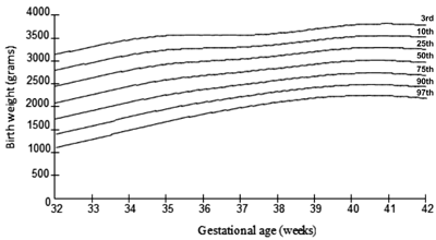
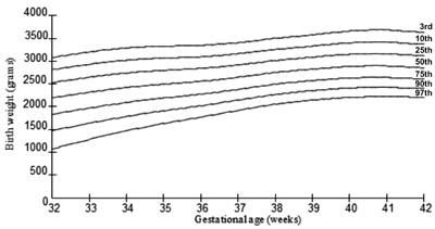
depicted in Table I-II, and Fig. 1,2. The
normality of Z scores calculated using the L, M and S values for
each gestational age was confirmed by Normal Q-Q plots for both genders
and gestational age categories within each gender.
TABLE I Centile Values for Males (n=8311)
|
Centile values* |
|
Age in weeks |
N |
3rd
|
10th
|
25th
|
50th
|
75th
|
90th
|
97th
|
|
32 |
53 |
1125.384 |
1418.11 |
1727.398 |
2084.334 |
2453.804 |
2796.073 |
3142.263 |
|
33 |
79 |
1307.635 |
1601.246 |
1909.976 |
2264.929 |
2631.248 |
2969.843 |
3311.737 |
|
34 |
120 |
1503.045 |
1793.081 |
2096.685 |
2444.482 |
2802.354 |
3132.4 |
3465.081 |
|
35 |
218 |
1691.319 |
1969.419 |
2259.296 |
2590.198 |
2929.677 |
3242.041 |
3556.329 |
|
36 |
361 |
1852.737 |
2110.664 |
2378.467 |
2683.149 |
2994.827 |
3280.956 |
3568.32 |
|
37 |
707 |
1994.273 |
2232.185 |
2478.42 |
2757.777 |
3042.841 |
3304.013 |
3565.884 |
|
38 |
1473 |
2117.048 |
2343.623 |
2577.654 |
2842.682 |
3112.685 |
3359.728 |
3607.16 |
|
39 |
1967 |
2205.521 |
2432.892 |
2667.591 |
2933.219 |
3203.687 |
3451.045 |
3698.701 |
|
40 |
1911 |
2254.88 |
2488.435 |
2729.537 |
3002.431 |
3280.316 |
3534.471 |
3788.945 |
|
41 |
1134 |
2251.521 |
2489.452 |
2735.158 |
3013.352 |
3296.714 |
3555.942 |
3815.545 |
|
42 |
288 |
2200.339 |
2441.596 |
2690.899 |
2973.335 |
3261.173 |
3524.612 |
3788.529 |
Table II Centile Values for Females (n=7683)
|
Centile values |
|
Age in weeks |
N |
3rd |
10th
|
25th
|
50th
|
75th
|
90th
|
97th
|
|
32 |
27 |
1071.636 |
1475.614 |
1835.385 |
2198.258 |
2533.816 |
2818.151 |
3085.657 |
|
33 |
54 |
1282.052 |
1637.941 |
1971.751 |
2320.082 |
2650.357 |
2935.297 |
3207.051 |
|
34 |
102 |
1471.989 |
1788.05 |
2095.316 |
2424.744 |
2743.945 |
3023.918 |
3294.427 |
|
35 |
174 |
1633.286 |
1913.546 |
2193.013 |
2499.096 |
2801.184 |
3070.089 |
3333.063 |
|
36 |
271 |
1774.326 |
2024.39 |
2278.336 |
2561.137 |
2844.551 |
3100.108 |
3352.778 |
|
37 |
535 |
1919.274 |
2148.231 |
2383.89 |
2649.787 |
2919.664 |
3165.744 |
3411.443 |
|
38 |
1163 |
2057.021 |
2273.012 |
2497.713 |
2754.026 |
3017.052 |
3259.297 |
3503.364 |
|
39 |
1892 |
2147.135 |
2355.66 |
2574.738 |
2827.229 |
3089.107 |
3332.698 |
3580.371 |
|
40 |
27 |
2206.053 |
2412.441 |
2631.489 |
2886.691 |
3154.404 |
3406.103 |
3664.593 |
|
41 |
1207 |
2227.793 |
2428.827 |
2644.25 |
2897.872 |
3166.935 |
3422.661 |
3688.011 |
|
42 |
299 |
2212.253 |
2403.482 |
2610.16 |
2855.84 |
3119.26 |
3372.265 |
3637.491 |
 |
|
Fig. 1 Birth weight centiles for males
- 3rd, 10th, 25th, 50th, 75th, 90th, 97th
|
 |
|
Fig. 2 Birth weight centiles for females - 3 rd,
10th, 25th, 50th, 75th, 90th, 97th
|
While the low birth weight proportion was 14%, the
proportion of small for gestational age was 8.1%. The median birth
weights of the study population were compared to the median birth
weights of US whites as well as UK white and Asian newborns (Web Fig.
I) [12,13].
Mortality: Mortality data analyzed for the years
2003 to 2012 for children for whom follow up data was available showed
that there were 207 deaths within the first two months among 14557 live
born singleton births, giving an under-two month mortality rate of
14/1000 live births. Of these 207 deaths, 146 (70%) occurred among the
children born to women without risk factors as included in the current
study. A comparison of mortality rates between children with lower birth
weight centiles and those in higher birth weight centile groups (
Web Table I) showed higher mortality at extremes
of birth weight.
Discussion
Previous Indian growth charts have been from tertiary
centres [4,5], which are often referral centres for those with antenatal
complications. However, the WHO growth standards provides median birth
weights for both sexes, for children born at term to healthy non-smoking
mothers [14], which can be taken as possibly representing ideal
birthweights.
The sex-specific growth charts produced by the
present study are descriptive of singleton live births of rural
antenatal women with no major antenatal risk factors, good antenatal
care, a high rate of institutional deliveries and a low birth weight
rate of 15% similar to the rest of Tamil Nadu (17.2%), but lower than
that of the entire country (22%). However, these growth charts are not
suitable as standards as the population is not the ideal one required
for the creation of standards.
Compared to the median birth weight for boys of 3.3
kg (3 rd centile 2.1 kg, 97th
centile 5 kg) in the WHO Child Growth Standards 2006 [14], where
healthy, socioeconomically well off mothers were included, in our study
males born at 40 weeks had a lower median birth weight of 3 kg (3rd
centile 2.25 kg, 97th
centile 3.8 kg). Similarly the median birth weight of females in the WHO
Growth standards was 3.2 kg (3rd
centile 2 kg, 97th centile
4.8 kg) which was higher than that of females in our study (median 2.9
kg, 3rd centile 2.2 kg, 97th
centile 3.6 kg). Comparison with birth weight centiles of the US and the
UK showed that while the weights of preterms were comparable to US
births, there was obvious faltering of the current study’s birth weights
at term, as compared to US whites, UK whites as well as south Asians in
the UK. A recent study from a tertiary centre in Andhra Pradesh also
showed lower birth weights as compared to international standards [5].
Although data regarding nutritional status by daily
caloric intake of this population was not recorded over the years, while
a previous survey from Tamil Nadu [15] showed that the median intake of
daily calories by pregnant women was below the recommended allowance.
The average height of the study women of 155 cm [6] was higher than the
national average of 152 cm, [16] but lower than that of developed
countries [17]. The shorter stature of Indian women while partly
explained by genetic variation, may also be due to under nutrition, as
even in developed countries, 20% of the variation in height is thought
to be due to environmental factors, with this proportion expected to be
higher for less developed countries [18].
The difference between the birth weights in the WHO
growth standards and our data illustrates that rural antenatal women
even without major antenatal risk factors had children with lower birth
weights. While major risk factors for low birth weight have been
excluded, low caloric intake remains a possibility which needs to be
further studied.
The mortality data highlighted the predictive value
of the birthweight centiles showing a much higher mortality among
children with birth weights below the 3 rd
centile as well as above the 97th.
This was interesting considering that the 97th
centile value in our data for e.g. 3.8 kg for males was below the
85th centile of the WHO
growth standards, weight for age for boys of 3.9 kg. Thus the mortality
experience of our neonates increases from birth weight centiles far
below the 97th centile of
the WHO growth standards (4.3 kg for boys), at weights which would be
considered normal by the WHO growth standards. This pattern of a J
shaped mortality curve with higher mortality at extremes of birth weight
[20] which is an established
phenomenon was also reflected in this study population although the
absolute values for birth weight seem to be situated to the left of the
widely accepted WHO growth standards.
A limitation of the data was that gestational age was
based mostly on last menstrual period, as performing ultrasound scans
for dating has not been a routine practice in rural areas. However, we
have attempted to reduce the error by removing birth weight outliers
(abnormally high/low birth weights for a gestation) and restricting to
an upper gestation of 42 weeks. Although we excluded mothers with known
antenatal risk factors affecting birth weight, it is possible that
undiagnosed risk factors for e.g. undiagnosed gestational
diabetes could have contributed to mortality among those with high birth
weights. Children with anomalies were also not excluded as this
information was not available.
The study findings raise the possibility that
increase in birthweights to match the WHO standards alone may not reduce
infant mortality in rural India as birthweights seem to be following a
different norm. While increase in birth weights would be of use in
preventing deaths in those in the lowest birth weight categories it may
not be necessary or even be harmful beyond a certain limit in its effect
on neonatal mortality. Comprehensive measures are needed to address both
outcomes for children with higher but so-called normal birth weights,
along with attempts to decrease low birth weights.
Contributors: All the authors have written,
contributed, designed and approved the study.
Funding: None; Competing interests: None
stated.
|
What is Already Known?
• Birth weight centile charts from tertiary
centers in India are available.
What This Study Adds?
• Birth weight centile curves based on
healthy pregnancies in a defined geographical region according
to sex.
• Under two month mortality rates of children with birth
weights both below the 3rd centile and above the 97th centile
were higher than those with normal birth weights (between 3rd
and 97th centiles) although the median birth weights were lower
than international standards.
|
References
1. Bonellie S, Chalmers J, Gray R, Greer I, Jarvis S,
Williams C. Centile charts for birthweight for gestational age for
Scottish singleton births. BMC Pregnancy Childbirth. 2008;8:5.
2. Seaton SE, Yadav KD, Field DJ, Khunti K, Manktelow
BN. Birthweight centile charts for South Asian infants born in the UK.
Neonatology. 2011;100:398-403.
3. Kierans WJ, Joseph KS, Luo ZC, Platt R, Wilkins R,
Kramer MS. Does one size fit all? The case for ethnic-specific standards
of fetal growth. BMC Pregnancy Childbirth. 2008; 8:1.
4. Mathai M, Jacob S, Karthikeyan NG. Birthweight
standards for south Indian babies. Indian Pediatr. 1993;33:203-9.
5. Kandraju H, Agrawal S, Geetha K, Sujatha L,
Subramanian S, Murki S. Gestational age-specific centile charts for
anthropometry at birth for South Indian infants. Indian Pediatr.
2012;49:199-202.
6. George K, Prasad J, Singh D, Minz S, Albert DS,
Muliyil J, et al. Perinatal outcomes in a South Asian setting
with high rates of low birth weight. BMC Pregnancy Childbirth. 2009 Feb
9;9:5.
7. Exploratory data analysis. J W Tukey.
Addison-Wesley;1977.
8. Francis MR, Rakesh PS, Mohan VR, Balraj V, George
K. Examining spatial patterns in the distribution of low birth weight
babies in Southern India- the role of maternal, socio-economic and
environmental factors. Int J Biol Med Res. 2012; 3:1255-59.
9. Cole TJ. Fitting smoothed centile curves to
reference data. J Royal Stat Soc. 1988;151:385-418.
10. Cole TJ. The use and construction of
anthropometric growth reference standards. Nutr Res Rev. 1993;6:19-50.
11. Cole TJ, Green PJ. Smoothing reference centile
curves: the LMS method and penalized likelihood. Stat Med.
1992;11:1305-19.
12. Oken E, Kleinman KP, Rich-Edwards J, Gillman MW.
A nearly continuous measure of birth weight for gestational age using a
United States national reference. BMC Pediatr. 2003;3:6.
13. WHO child growth standards:
length/height-for-age, weight-for-age, weight-for-length,
weight-for-height and body mass index-for-age: methods and development.
Geneva: World Health Organization, 2006. Available from:
http://www.who.int/childgrowth/standards/Technical_ report.pdf. Accessed
24 May, 2012.
14. WHO child growth standards, weight-for- age.
Available from:
http://www.who.int/childgrowth/standards/weight_for_age/en/index.html.
Accessed 24 May, 2012.
15. Diet and Nutritional Status of Rural Population.
National Nutrition Monitoring Bureau. NNMB Technical Report 21.
Available from: http://www.nnmbindia.org/NNMBREPORT2001-web.pdf.
Accessed 24 May, 2012.
16. Mamidi RS, Kulkarni B, Singh A. Secular trends in
height in different states of India in relation to socioeconomic
characteristics and dietary intakes. Food Nutr Bull. 2011;32:23-34.
17. Ogden CL, Fryar CD, Carroll MD, Flegal KM. Mean
body weight, height, and body mass index, United States 1960-2002. Adv
Data. 2004;(347):1-17.
18. Silventoinen K. Determinants of variation in
adult body height. J Biosoc Sci. 2003;35:263-85.
19. Wilcox AJ, Russell IT. Birthweight and perinatal
mortality: II. On weight-specific mortality. Int J Epidemiol
1983;12:319-25.
|
|
|
 |
|
