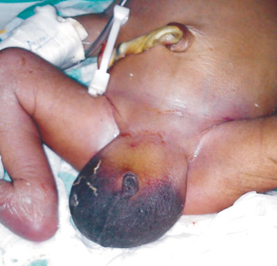
A term male neonate weighing 2640 grams, born after 37 weeks of
gestation by vaginal delivery had enlarged scrotum at birth. Antenatal
period was complicated by polyhydramnios; ultrasonography done at 34
weeks had revealed dilated stomach and bowel loops. The scrotal swelling
was soft, fluctuant, non-tender, non-reducible, bright on
transillumination with bilateral testes being palpated separately (Fig.
1). He gradually developed abdominal distension, and bilious
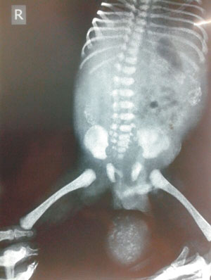
aspirates by end of day 1 of life. Plain X-ray of the abdomen
showed scattered peritoneal and scrotal calcifications (Fig. 2).
Diagnosis of intestinal obstruction with Meconium periorchitis was made.
Explaratory laporotomy revealed Ileal atresia that was treated by
resection and primary anastomosis. Scrotal swelling resolved completely
by 3 months on follow-up.
 |
|
Fig. 1 Huge scrotal swelling
with penis buried in scrotal mass.
|
 |
|
Fig. 2 X-ray of abdomen showing
peritoneal and scrotal calcifications.
|
Meconium periorchitis occurs when meonium in the
peritoneal cavity reaches the paratesticular tissue through patent
processus vaginalis, and incites intense inflammatory reaction.
Typically, the scrotal swelling is mistaken for hydrocele at birth but
the mass progressively hardens due to calcification. Isolated
calcifications in the scrotum without peritoneal involvement are seen in
neoplasms such as teratoma, rhabdomyosarcoma, metastatic neuroblastoma,
lymphoma and pseudotumors. The visualization of the normal testicle on
ultrasonography differentiates meconium periorchitis from scrotal tumors.
Epididymal inflammation, torsion testis and scrotal abscess are other
testicular masses at this age; these present as painful scrotal mass.


