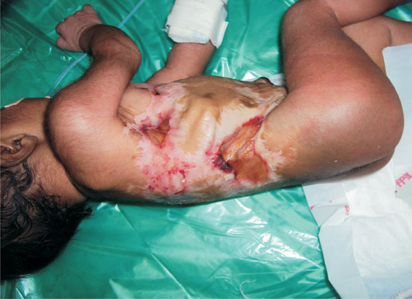
A girl weighing 2320 grams was delivered at 38 weeks to a mother with
antenatal history of chickenpox in the fourteenth week of gestation. At
birth, baby had cicatricial lesions with areas of induration and
hypopigmentation on the torso, causing deformity of underlying rib cage
(Fig. 1). The limbs were spared. She had bilateral
chorioretinal atrophy with cataract in the left eye. Her sensorium was
altered, and she had poor respiratory efforts requiring mechanical
ventilation. Computed tomography of brain revealed bilateral thalamic
calcifications and mild cerebral atrophy. Despite intensive care, baby
died at fifty hours of life.
 |
| |
The risk of congenital varicella syndrome (CVS) after
maternal varicella in the first 20 weeks of pregnancy is approximately
0.4 - 2%. Cicatricial lesions, neurological defects, ophthalmological
manifestations and limb-shortening defects with muscular hypoplasia are
the major clinical features in CVS. An important differential diagnosis
for cicatricial lesions in a neonate is intrauterine Herpes Simplex
Virus infection (distributed in dermatomal fashion but presence of
active lesions, hypo- and hyperpigmentation, aplasia cutis, and/or an
erythematous macular exanthema).


