Intramuscular hemangiomas are rare tumors
constituting a mere 0.8% of all hemangiomas(1,2). They deserve attention
not only because of their rarity but also because of their invariably
confusing clinical presentation as well as intriguing etio-pathogenesis.
Case Report
A 12-year-old girl presented with a painful swelling
in the left forearm. There was history of significant trauma to the left
forearm, at the age of 6 years. Following the injury, she was initially
taken to a traditional bonesetter who splinted and immobilized the limb
for 4 weeks. The child was asymptomatic and had no movement deficits on
splint removal. Over the next few years, she gradually developed a
painful swelling in the left forearm and began finding it increasingly
difficult to use the affected hand. The swelling was diffuse and slow
growing and was not associated with any constitutional symptoms like
fever or weight loss.
Local examination suggested a diffuse intramuscular
mass in the flexor compart-ment. It was warm, tender and boggy. The
swelling was neither compressible nor pulsatile and no bruit could be
heard on auscultation. The wrist and fingers were held in flexion and
passive extension was grossly limited and very painful. Active flexion
of the fingers and wrist was also restricted and painful. Passive
flexion of the wrist permitted some extension of the fingers at the
interphalangeal joints (i.e., a positive Volkmann’s sign).
However, as the patient had severe pain on both active extension and
flexion, the sign was considered false positive and not much
significance was attached to it at this stage. A clinical diagnosis of
chronic flexor tenosynovitis was made. Radiography revealed a soft
tissue mass with multiple calcific spots and the following differential
diagnoses were considered: tuber-culous flexor tenosynovitis (with
calcified melon seed bodies) or an organized hematoma (with metastatic
calcification).
Surgery revealed a multiloculated purple-red mass
that had completely involved the flexor digitorum superficialis (FDS).
Further, the mass had invaded into the substance of the median nerve at
the wrist and had enveloped all other muscles, tendons and neurovascular
structures in the distal three-fourths of the forearm. The mass was not
compressible even intraoperatively and no feeder vessel could be
demonstrated. The median nerve required an internal neurolysis as the
mass had insinuated between its fascicles. It was possible to dissect
the mass away from all other musculo-tendinous and neurovascular
structures in the forearm except the FDS, whose belly was excised along
with the tumour. As there was no reason at this stage to suspect any
muscle necrosis, no specific attempt was made to explore the flexor
profundus or flexor polices longus (FPL) but the muscle looked healthy
on gross appearance. Post excision, the remaining long flexor tendons (i.e.,
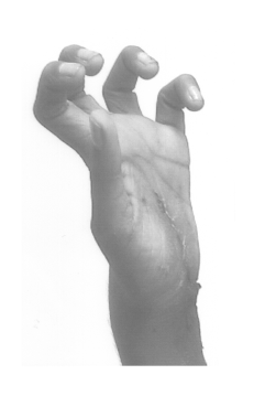
FDP and FPL) were found contracted with a persisting Volkmann’s sign (Fig.
1).
 |
|
Fig. 1. Early Postoperative picture showing
Volkmann’s sign. |
Histopathology of the excised mass revealed muscle
tissue with a well-defined lesion comprising closely packed thin walled
capillaries, areas of hemorrhage and foci of calcification. The final
diagnosis was an intramuscular capillary haemangioma.
Electrophysiological studies of the ulnar and median
nerves confirmed no damage to innervation during dissection of the
tumor. Post-operatively, the patient underwent intensive physiotherapy
for stretching of the contracted tendons and for restoration of finger
flexor power as she was left only with the flexor profundus. Full
functional restoration was achieved about 8 months after surgery.
Discussion
The exact cause of the intramuscular hemangioma has
been something of an enigma. Some authors feel that it is a con-genital
neoplasm while others have reported a previous history of trauma(3).
Allen and Enzinger(2) have reported trauma to be the cause in 5%
patients in their series. A definite history of trauma was available
even in the present case.
Virtually, every report of the intra-muscular
hemangioma in literature stresses its atypical clinical presentation. It
is usually seen as a slow growing mass that may or may not be painful.
Features characteristic of vascular tumors like pulsation, thrill or
bruit are generally absent and hence, the condition is rarely diagnosed
pre-operatively(2). Fergusson(4) reports that the intramuscular
haemangioma has been clinically misdiag-nosed in more than 90% of cases.
Pain is a common symptom, contra-dictory to that expected in a benign
neoplasm. This has been attributed to the peculiar association of
abnormal blood vessels with nerve fibers as well as significant levels
of Substance P in these tumors(5,6). The current patient presented with
all features suggesting an inflammatory process. Most significantly, she
had severe pain on direct pressure over the mass as well as on resisted
flexion or passive stretching of the fingers. This led to a mistaken
clinical diagnosis of flexor tenosynovitis.
Histopathologically, the tumor can be classified into
three types, capillary, caver-nous and mixed, based on the predominant
vascular pattern(2). The present case was of the capillary type.
Management of intra-muscular hemangioma has evolved over time.
Spontaneous resolution has not been known to occur(3). Radiotherapy,
cryotherapy and embolization have been found to be largely
ineffective(3,4). Rogalski et al.(5) in their series of 41
patients could not find a feeder vessel large enough for embolization.
There was no feeder vessel evident in the present case as well. Total
excision, often requiring en block resection of the tumor along
with the involved muscle is the universally recommended treatment and
was the method employed in the current case.
Various complications have been reported in
intramuscular hemangiomata, but so far, contracture of adjacent
uninfiltrated muscles has not been reported. The current case appears to
display this unique complication. The features of contracture were very
much like that seen in Volkmann’s ischaemic contracture (VIC) and
involved the FDP and FPL. It is tempting to speculate whether the
contracture occurred as a result of chronic muscle ischemia. Factors
which contribute to this line of thought are: (i) The Volkmann’s
sign persisted even after excision of the tumor; (ii) the tumour
had invaded the FDS only; (iii) more superficial muscles like the
wrist flexors or pronator teres were not involved (iv) The flexor
profundus and the flexor pollicis longus were contracted. These are
typically the muscles to be affected in the localized type of VIC. The
contracted muscles were enveloped but not infiltrated by tumor tissue
and there was a clear plane of dissection between the tumor and
enveloped muscles. This suggests a cause of contracture other than tumor
induced necrosis or fibrosis(7). Robinson et al. have reported a
Substance-P and CGRP, which are found in intramuscular haemangiomata,
are known to divert blood away from muscle fibers into the surrounding
connective tissue. It is debatable whether this is confined only to the
infiltrated muscle or can exert influence on surrounding muscles, thus
contributing to ischaemia and fibrosis. Rogalski et al.(5) have
reported compartment syndrome following Intramuscular hemangioma in the
forearm. Other authors have reported compartment syndrome following
other tumors or mass lesions in closed osteofascial compart-ments(8,9).
Anderson and Chandi(10) have reported contracture of the flexor
profundus following cysticercosis. Though cysticercus infiltrates of
muscles are usually asympto-matic, the excised muscle mass was pale and
fibrotic. They speculated that there might have been a segmental
vascular compromise of the muscle, which led to fibrosis and
contracture. The presence of a steadily grow-ing mass in closed
osteofacial compartment of the forearm in this case, had in all
probability caused a chronic compartment syndrome. Whether this led to
ischemia is worthy of consideration. Following surgery, the patient
underwent an intensive physiotherapy program comprising stretching and
FDP and FPL strengthening exercise. By 32 weeks post-excision, the
patient had recovered full range of movement as well as grip strength
comparable to the opposite hand.
Funding: None.
Competing interests: None stated.
