|
|
|
Indian Pediatr 2016;53:
525-527 |
 |
Blue Rubber Bleb Nevus Syndrome with Musculo-skeletal
Involvement and Pulmonary Stenosis
|
|
A Singal, S Vohra, R Sharma and *S Bhatt
From Departments of Dermatology and STD, and *Radiology, University
College of Medical Sciences and GTB Hospital (University of Delhi),
Dilshad Garden, Delhi, India.
Correspondence to: Dr (Prof) Archana Singal, University College of
Medical Sciences and GTB Hospital,
Delhi 110 095, India.
Email:
[email protected]
Received: September 12, 2015;
Initial review; October 26, 2015;
Accepted: February 13, 2016.
|
|
Background: Blue rubber bleb nevus syndrome is a rare
clinical entity. Case characteristics: A 13-year-old Indian boy
presented with characteristic cutaneous lesions, gastrointestinal
malformations, skeletal involvement and pulmonary stenosis.
Observation: Diagnosis was confirmed on skin biopsy, radiographic
evaluation, colonoscopy and echocardiography. Echocardiography revealed
pulmonary stenosis, an association hitherto undescribed. Message:
Detailed evaluation in a patient of blue rubber bleb nerves syndrome is
mandatory.
Keywords: Hemangioma, Pulmonary stenosis,
Vascular malformations.
|
|
B
lue rubber bleb nevus syndrome (BRBNS, Bean
syndrome) is a rare disorder with venous malformations involving skin
and other organs especially gastrointestinal tract. William Bennet Bean
in 1958 coined the term
‘blue ruber bleb syndrome’ on the basis of bluish color of the nevi and
their rubbery consistency [1]. Cutaneous vascular malformations,
characterized by multiple, asymptomatic, bluish and rubbery blebs are
located on trunk and upper extremities. Gastrointestinal (GI) lesions
are commonly located in the small intestine causing occult and chronic
bleeding leading to iron deficiency anemia [2]. Herein, we describe a
case of BRBNS in an adolescent boy with prominent musculo-skeletal
involvement and pulmonary stenosis.
Case Report
A 13-year-old boy presented with multiple blue raised
skin lesions all over the body. Starting in the first year of life the
lesions first appeared on the nape of the neck and gradually increased
in number and size. He denied history of seizures, headache, vision/
hearing defect, dyspnea, dysphagia, GI bleed or pain abdomen. Physical
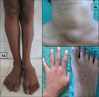
examination revealed moderate pallor and an eversion deformity of the
left foot (Fig. 1a). The length and girth of the left leg
was decreased as compared to the right side (82 vs 87 cms when
measured between anterior superior iliac spine and lateral malleolus and
21cm vs 24 cm measured at the junction of middle and lower one
third of leg) leading to short limb gait along with the scoliosis of
dorso-lumbar spine.
 |
|
Fig. 1 Eversion deformity of the left
foot; (a) Bluish papule and subcutaneous swellings around neck;
(b) and lesions over hands and feet (c).
|
Multiple, discrete, skin and blue coloured smooth
surfaced papules, nodules and subcutaneous swellings (0.5 - 4cm size)
were distributed over the right lower eye lid, around the neck, sternal
notch, retroauricular regions and fewer lesions over trunk, bilateral
upper and lower extremities (Fig. 1). These lesions were
soft, partially to completely compressible, non-pulsatile and non-tender
without ulceration and haemorrhage. Except for moderate microcytic
hypochromic anemia (hemoglobin 8 g/dL), serum chemistry and coagulation
study were normal. Stool for occult blood was negative.
Skin biopsy from the lesion showed presence of
ectatic channels in the dermis lined by endothelium. Colour Doppler
study confirmed the low flow nature of lesions. Radiograph of the dorso-lumbar
spine revealed mild scoliosis with concavity towards the left in the
dorsal and towards right in the visualized lumbar spine). Scoliosis
occurred as a compensatory phenomenon due to shortening of the left leg.
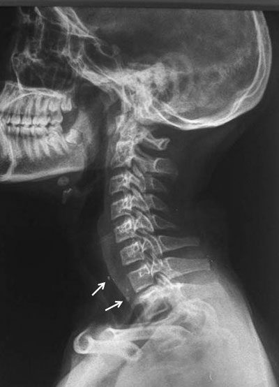
Radiograph of the neck lateral view showed multiple rounded
radio-densities consistent with phleboliths (Fig. 2).
X-rays of left lower limb revealed cortical irregularities, bowing of
the fibula, multiple soft tissue calcifications and generalized
osteopenia. There was restricted growth of distal lateral epiphysis of
left tibia leading to eversion deformity of the left foot Magnetic
resonance imaging brain, ultrasonography (USG) thyroid and eyes, upper
gestrointestinal (GI) endoscopy and computed tomography scans of abdomen
and pelvis were normal. Colonoscopy showed numerous, 3-5 mm bluish
nodules studded on the mucosal surface of rectum, sigmoid and descending
colon. Echocardiography revealed pulmonary stenosis.
 |
|
Fig. 2 Radiograph of the neck lateral
view shows multiple rounded radio-densities (arrow) consistent
with phleboliths.
|
The child was started on oral hematinics for anemia.
Parents were counselled regarding the possible gastrointestinal
complications and importance of periodic gastroenterology and cardiology
follow up.
Discussion
BRBNS is a rare disorder characterized by multiple
venous malformations in the skin, gastrointestinal tract and less often
in the central nervous system, musculoskeletal system, thyroid,
parotids, eyes, oral cavity, kidney, liver, and bladder [3]. Diagnosis
of BRBNS is based on the presence of characteristic asymptomatic
cutaneous venous malformations that are usually present at birth or
appear in early life while other organ involvement appears later, as
such in our case. Lesions are preferentially located on the upper limbs,
trunk, and perineum. Venous malformations of the lower limbs are unusual
and are often associated with significant orthopedic involvement as seen
in present case [4].
Intestinal lesions cause chronic bleeding and rarely
present with acute hematemesis or melena. Rarely, complications like
intussusception, volvulus, infarction or hemorrhage can occur. There are
few reports of BRBNS with associated cardiac defects like atrial and
ventricular septal defects and pulmonary hypertension secondary to
thromboembolic events [5,6]. Our patient had pulmonary stenosis, an
association hitherto undescribed.
Orthopedic manifestations include skeletal bowing,
pathological fractures, articular problems and bony overgrowth that
arise as a result of pressure effects from adjacent vascular lesions.
Presence of scoliosis, pectus excavatum, and congenital dysplasia of hip
and club foot in a new born with BRBNS has been described [7]. However,
in the present case, skeletal anomalies were late in onset and included
dorso-lumbar scoliosis, bowing of fibula and eversion deformity of foot.
Therapeutic options in BRBNS depend upon the clinical
manifestations. Cutaneous lesions require treatment when they are
cosmetically unacceptable or functionally troublesome. Ruby, argon, and
carbon-dioxide laser treatments, electrodessication, scalpel excision,
and injection sclerotherapy have been tried. Medical treatment for
gastrointestinal lesions includes oral hematinics for anemia, interferon
gamma, octreotide and sirolimus [8,9]. In addition, endoscopic
sclerotherapy, surgical bowel resection and photocoagulation have been
tried. For orthopedic abnormalities, physiotherapy, serial plaster cast
correction, braces or surgical excision have been used. The prognosis
varies with the extent of visceral organ involvement and most patients
tend to have normal life span.
We conclude that a detailed systemic evaluation is
recommended in patients with BRBNS to look for asymptomatic organ
involvement and advice on regular follow-up and early institution of
appropriate treatment.
Contributors: AS: Patient management and
diagnosis; SV: Patient management, searching related literature and
writing the manuscript; RS: literature review and writing the
manuscript; SB: Radiographic evaluation and diagnosis.
Funding: None; Competing interests: None
stated.
References
1. Bean WB. Vascular spiders and related lesions of
the skin. IL, Springfield.Thomas, 1958.
2. Moodley M, Ramdial P. Blue rubber bleb nevus
syndrome: case report and review of literature. Pediatrics.
1993;92:160-2.
3. Haiping P, Guoqing L, Zhongshu Y. Blue rubber bleb
naevus syndrome. Eur J Surg. 2001;167: 628-30.
4. McCarthy JC, Goldberg MJ, Zimbler S. Orthopedic
dysfunction in blue rubber bleb nevus syndrome. J Bone Joint Surg Am.
1982;64:280-3.
5. Aroor S, Varma C, Mundkur SC. Blue rubber-bleb
vevus syndrome which was associated with an atrial septal defect: A case
report. J Clin Diagn Res. 2012;6:1566-7.
6. Giordano C, Battagliese A, di Gioia CR, Campagna
D, Benedetti F, Travaglini C, et al. Blue rubber bleb nevus
syndrome and pulmonary hypertension: An unusual association. Cardiovasc
Pathol. 2004;13:317-22.
7. Tzoufi MS, Sixlimiri P, Nakou I, Argyropoulou MI,
Stefanidis CJ, Siamopoulou-Mavridou A. Blue rubber bleb nevus syndrome
with simultaneous neurological and skeletal involvement. Eur J Pediatr.
2008;167:897-901.
8. Gonzalez D, Elizondo BJ, Haslag S, Buchanan G,
Burdick JS, Guzzetta PC, et al. Chronic subcutaneous octreotide
decreases gastrointestinal blood loss in blue rubber-bleb nevus
syndrome. J Pediatr Gastroenterol Nutr. 2001;33:183-8.
9. Yuksekkaya H, Ozbek O, Keser M, Toy H. Blue rubber
bleb nevus syndrome: successful treatment with sirolimus. Pediatrics.
2012;129:1080-4.
|
|
|
 |
|
