|
|
|
Indian Pediatr 2013;50: 623 |
 |
Chronic Bullous Disease of Childhood
|
|
Rashmi Mahajan, Priyank Shah and Sheela Bharani
Department of Dermatology, SBKS Medical College and
Research Centre, Gujarat, India.
Email: [email protected]
|
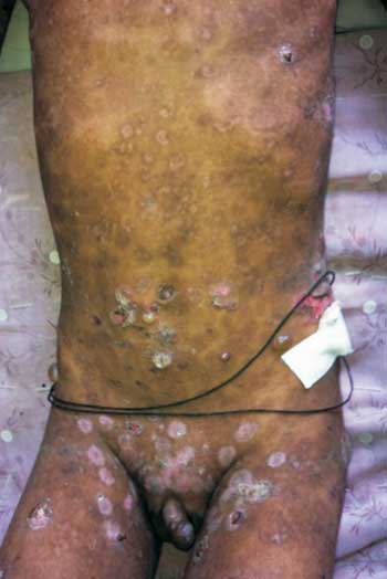
A 9-year-old boy presented with itching and bulla formation
since the last 3 years. The lesions commenced over the
abdomen, scalp and then became generalized. Past perinatal
and drug history were uneventful. On examination, tense
bullae (0.5 cm to 3.5 cm in diameter) were present all over
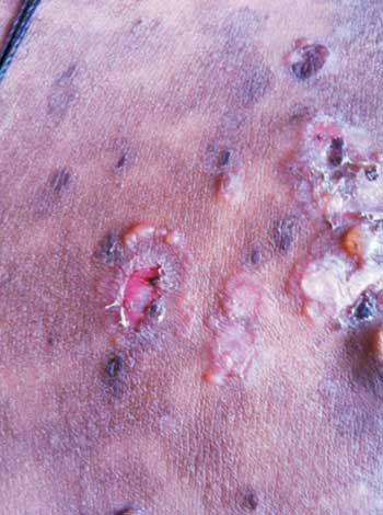
the body (Fig. 1). Bullae arranged in an
annular pattern were seen in perineal region (string of
pearl’s sign) (Fig. 2). Bulla spread and
Nickolsky sign were negative. Tzanck smear was negative for
acantholytic cells. Based on above findings, a differential
diagnosis of chronic bullous diseases of childhood,
childhood bullous pemphigoid and dermatitis herpetiformis
were considered. Skin biopsy revealed bulla formation
between basal layer of epidermis and dermis, papillary
dermal edema, dilated blood vessels with mild perivascular
inflammatory infiltrate comprising of lymphocytes,
neutrophils and eosinophils. Direct immunofluorescence test
(for which patient was not affording) is confirmatory for
Chronic bullous disease of childhood, demonstrating linear
deposition of IgA at basement membrane zone. While this
disease has been defined on the basis of its unique
immunopathology and occurs in both adults and children,
however, in children the cutaneous features may be
clinically unique. The disease responds to dapsone at less
than 0.5 mg/kg/day, with regular monitoring of hemoglobin
levels. Spontaneous remission occurs after 3-6years.
 |
 |
|
Fig.1 Bullae all over body.
|
Fig.2 String of pearl sign.
|
|
|
|
 |
|
