|
|
|
Indian Pediatr 2018;55: 601- 602 |
 |
Choroid Osteoma in Schimmelpenning-Feuerstein-Mims
Syndrome
|
|
Vasanthan Tanigasalam 1,
B Vishnu Bhat1,
Sharmila Manivannan2,
Malathi Munisamy3
and
Swapnil Parchand4
From Departments of 1Neonatology, 2Pediatrics,
3Dermatology, and 4Ophthalmology, Jawaharlal
Institute of Postgraduate Medical Education and Research (JIPMER),
Puducherry, India.
Correspondence to: Vasanthan Tanigasalam, Department
of Neonato-logy, JIPMER, Puducherry 605 006, India.
Email:
[email protected]
Received: May 09, 2017;
Initial review: June 21, 2017;
Accepted: March 10, 2018.
|
Background: Schimmelpenning syndrome is a multisystem disorder.
Case characteristics: A term female neonate with sebaceous nevi of
the face had choroid osteoma of the right eye. Observation: At
one month of age, the infant was observed to have choroidal
neovascularization that was successfully treated with laser
photo-coagulation and anti-VEGF. Message: Choroid osteoma and
neovascularization are rare associations of Schimmelpenning syndrome,
and should be screened for and managed early.
Keywords: Choristoma, Management, Retinal coloboma, Screening.
|
|
S
chimmelpenning-Feuerstein-Mims Syndrome is a rare
multisystem disorder characterized by sebaceous nevus associated with
other abnormalities of the brain, eyes, and bones. Sebaceous nevi
clinically presents as hairless hypopigmented to yellowish circumscribed
plaques with a tendency to become more verrucous with puberty. These
nevi demonstrate hyperplasia of the epidermis, immature hair follicles,
and irregularities of morphology and distribution of sebaceous glands
[1]. Sebaceous nevus when associated with multiple organ involvement is
the organoid nevus syndrome known as Schimmelpenning syndrome [2]. We
report the presence and successful management of choroidal osteoma
induced neovascularization in a neonate who had sebaceous nevi
associated with ophthalmologic and cardiovascular involvement.
Case Report
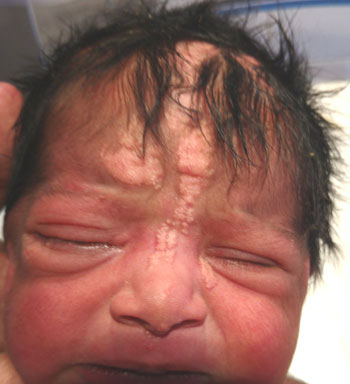
A term female neonate, born out of third degree
consanguineous marriage with an uneventful antenatal period, presented
with hypopigmented-to-yellowish plaques pathognomonic of sebaceous nevi
over the forehead and chin in a blaschloid distribution associated with
non-scarring alopecia of the scalp (Fig. 1).
Ophthalmologic examination demonstrated right corneal haziness with
vascularization. Slit lamp examination revealed a calcified lesion over
the bulbar conjunctiva with the possibility of choristoma of the
conjunctiva; there was no cataract. Fundus examination showed right
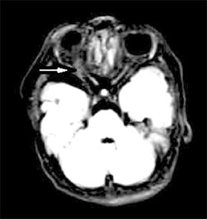
retinal coloboma. Contrast enhanced computed tomography revealed
calcified lesions of the right optic canal. Magnetic resonance imaging
was suggestive of right retinal coloboma associated with calcified
lesion around the optic nerve suggestive of choroid osteoma. The
echocardiogram revealed ventricular septal defect. There was no
genitourinary abnormality. Skeletal survey, and serum levels of calcium,
phosphorous, alkaline phosphatase, parathyroid hormone and 25-hydroxy
vitamin D were normal. The neonate was otherwise asymptomatic and
discharged on day 7 of life. On follow-up at one month of age, the
infant was observed to have choroidal neovascularization that was
treated with laser photocoagulation and intravitreal anti-VEGF (bevacizumab).
At four months of age, there was a gradual regression of choroidal
neovascularization, and preserved vision.
 |
|
Fig. 1 Hypopigmented-to-yellowish
hairless skin lesion suggestive of sebaceous nevi.
|
 |
|
Fig. 2 MRI showing right retinal
coloboma and choristoma (arrow).
|
Discussion
Schimmelpenning syndrome is characterized by
sebaceous nevi of Jadossohn with the eye, brain and skeletal defects.
Observed prevalence is 1 to 3 per 1000 live births. The occurrence of
the disease is sporadic due to postzygotic mutation. In 60-80% of the
cases, the skin lesions are confined to the scalp and face region, and
are associated with nonscarring alopecia of scalp [3].
Choristoma of the conjunctiva, coloboma of the eye,
epibulbar dermoid and scarring degeneration of the retina are the most
common ocular findings in this syndrome [4,5]. In this case, apart from
the common ocular findings like choristoma of the bulbar conjunctiva and
retinal coloboma, we observed an unusual calcified lesion suggestive of
choroid osteoma in the right eye. Choroid osteoma is a benign tumor of
mature bone replacing the choroid. Some consider it as choristoma in the
region of choroid [6]. Visual loss may result from atrophy of retinal
pigment epithelium overlying the osteoma, serous retinal detachment over
the osteoma, and choroidal neovascularisation. In the present case, the
neonate developed choroidal neovascularization that was successfully
treated with laser photocoagulation and intravitreal anti-VEGF (bevacizumab),
thus preserving the vision [7].
Neurological abnormalities observed in this syndrome
include hemi megalencephaly, corpus callosum agenesis, gyri
malformations and Dandy-Walker cyst. Skeletal defects observed are
vertebral defects, craniofacial defects, asymmetry of skull bones and
bone cysts. Other uncommon association includes coarctation of the
aorta, genitourinary abnormalities, and vitamin D resistant rickets.
There is a predisposition to develop benign skin tumors in the sebaceous
nevi [5]. The overall prognosis depends on the severity of associated
anomalies. In the present case, the neonate had ventricular septal
defect with no neurological and skeletal defects.
We conclude that choroid osteoma and choroidal
neovascularisation are rare ophthalmologic mani-festations of
Schimmelpenning syndrome which should be screened for so that initiation
of early treatment results in better visual outcome.
Contributors: VB, SM, MM and SP: case management
and supervision; VT: drafted the manuscript, which was revised and
approved by all authors.
Funding: None; Competing interest: None
stated.
References
1. Warnke PH, Hauschild A, Schimmelpenning GW,
Terheyden H, Sherry E, Springer IN. The sebaceous nevus as part of the
Schimmelpenning-Feuerstein-Mims syndrome - an obvious phacomatosis first
documented in 1927. J Cutan Pathol. 2003;30:470-2.
2. Happle R. The group of epidermal nevus syndromes
Part I. Well defined phenotypes. J Am Acad Dermatol. 2010;63:1-22; quiz
23-4.
3. Van de Warrenburg BP, van Gulik S, Renier WO,
Lammens M, Doelman JC. The linear naevus sebaceus syndrome. Clin Neurol
Neurosurg. 1998;100:126-32.
4. Shields JA, Shields CL, Eagle RC Jr, Arevalo JF,
DePotter P. Ocular manifestations of the organoid nevus syndrome.
Ophthalmology. 1997;104:549-57.
5. Insler MS, Davlin L. Ocular findings in linear
sebaceous naevus syndrome. Br J Ophthalmol. 1987;71:268-72.
6. Alameddine RM, Mansour AM, Kahtani E. Review of
choroidal osteomas. Middle East Afr J Ophthalmol. 2014;21:244-50.
7. Papastefanou VP, Pefkianaki M, Al Harby L, Arora
AK, Cohen VM, Andrews RM, et al. Intravitreal bevacizumab
monotherapy for choroidal neovascularization secondary to choroidal
osteoma. Eye (Lond). 2016;30:843-9.
|
|
|
 |
|
