|
|
|
Indian Pediatr 2021;58: 88-89 |
 |
Beneficial Response to Phosphate Lowering Therapy in
Normophosphatemic Tumoral Calcinosis
|
|
Sarayu Soumya,1 Nandini Prasad,2
Puthiyaveettil Khadar Jabbar,1 Sajid Hussain,3
Chellamma Jayakumari4 and Abilash Nair1*
From Departments of 1Endocrinology and
Metabolism, 3Orthopaedics, 4Internal Medicine,
Government Medical College; Thiruvananthapuram; 2Pediatrics,
Health Services Department,
Government of Kerala, Kerala, India.
Email:
[email protected]
|
|
Calcinosis cutis is a comprehensive terminology for disorders
characterised by deposition of calcium salts in the cutaneous and
subcutaneous tissue. Based on etiology, it can be classified into:
dystrophic, metastatic, idiopathic, iatrogenic, and calciphylaxis.
Dystrophic calcinosis occurs following a necrotic process unrelated to
serum calcium level. Metastatic calcification results from precipitation
of calcium salts due to high calcium or phosphorus levels. Calciphylaxis
denotes calcification of the small and medium-sized blood vessels, of
the dermis or subcutaneous tissue usually in the setting of renal
failure [1]. Tumoral calcinosis refers to a severe form of calcinosis
involving deeper tissues, especially around joints leading to limitation
of movement.
A 14-year-old boy presented with history of hard
swellings in the skin around multiple joints for past two years. Some of
them ulcerated to extrude chalky white material. There was restriction
of movements at knees, shoulders and elbows. He did not have history or
examination findings to suggest autoimmune connective tissue disorders,
pancreatic disease, chronic renal failure, malignancy, trauma, medical
intervention in the affected regions. His previous records showed normal
calcium and phosphorus levels, and he was not on calcium or vitamin D
supplementation. There was no family history of similar condition. He
had undergone excision of a large lesion from right axilla but did not
receive any oral or parenteral medications for treatment of lesions
before presenting to us.
There were multiple hard swellings involving the skin
and subcutaneous tissue around shoulder, elbow, hip, knee joint and neck
on both sides (Fig. 1a). Range of movement of all large joints
measured using a universal goniometer showed restriction of joint
movements which was maximum at right knee joint (50 degrees) and right
elbow (30 degrees) for extension. Restriction of movements at the knee
joints had resulted in limping. The physical examination was otherwise
unremarkable.
Blood counts, renal function, liver function
parameters were normal. Average serum calcium and phosphorus
level(fasting sample) after multiple measurements were normal (9.4 mg/dL
and 4.8mg/dL, respectively).Vitamin D insufficiency (25-hydroxy vitamin
D level 12ng/mL) and secondary hyper-parathyroidism (Serum PTH level 126
pg/mL) were present. Plasma levels of 1, 25- dihydroxy Vitamin D was
normal. Anti-nuclear antibody (ANA), Anti ds-DNA, Anti neutrophil
cytoplasmic antibody (ANCA), anti-centromere, anti scl-70 and U1RNP were
normal. Radiographic skeletal survey showed multilobulated ‘cloud-like’soft
tissue calcification around joints with normal joint morphology (Fig.1b,c).
Sonographically there were no renal and ureteric calculi or
nephrocalcinosis. He had undergone excision of a lesion from the axilla
before presenting to us, and the histopathology report had shown
multiple cysts filled with calcified deposits lined by histiocytes
consistent with tumoral calcinosis.
 |
|
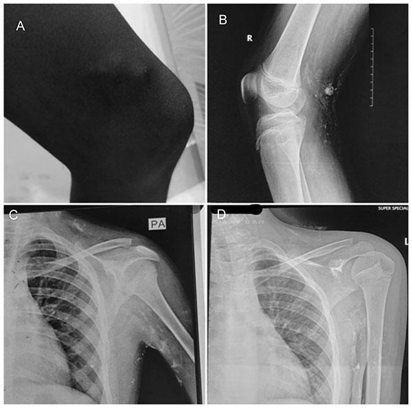
Fig. 1 (a) periarticular hard
swellings at right knee, (b) X-ray of right knee joint
showing multiple "cloud- like" periarticular calcifications, (c)
X-Ray showing periarticular calcifications at left axilla
and neck along platysma, (d) X-Ray of left axilla after 6
months of treatment with sevelamer and low phosphorus diet
showing decrease in extent of calcification at axilla and neck.
|
Differential diagnoses considered were disorders
which cause dystrophic calcification (like infection, inflammatory
processes, cutaneous neoplasm or connective tissue diseases), metastatic
calcification (like hypercalcemia or hyperphos-phatemia) and secondary
tumoral calcinosis due to chronic kidney disease. All these
differentials were effectively excluded by evaluation. Final diagnosis
of normophosphatemic idio-pathic tumoural calcinosis was made.
Multidisciplinary team including endocrinologist,
ortho-paedician, paediatrician and physiatrist decided to start
phosphate lowering medical management aiming to keep phos-phorus in the
low normal range, with graded physiotherapy and follow up closely after
obtaining patients consent. Along with a low phosphorus diet, sevelamer
at a dose of 400 mg twice daily was given orally. Surgical correction of
deformity was reserved for a later scenario if medical and physical
measures failed.
After 6 months of follow upon therapy, he reported
improvement in joint movements and limp had disappeared which were
confirmed on examination. X-rays showed decrease in the size of
calcifications at axilla, knee and neck (Fig. 1d), whereas the
lesion in the elbows did not increase in size. Smaller lesions have
disappeared also. During the one year of follow-up, while maintaining
serum phosphorus level between 3.3mg/dL and 3.5 mg/dL, he reports no new
skin lesions or movement restriction nor any adverse events related to
therapy.
Tumoral calcinosis is divided into hyperphosphatemic
(familial), normophosphatemic and secondary variants [2].
Hyperphosphatemic form is the most common. High phosphorus levels are
due to a reduced renal clearance due to decreased action of the
phosphaturic hormone fibroblast growth factor 23 (FGF23) which in
turn may be due to mutated FGF23 or an enzyme involved in
stabilization of wild type FGF23, or
a-klotho (the
cofactor for FGF23 action). Disorders like chronic renal failure
with secondary hyperparathyroidism and hyper-vitaminosis D cause the
secondary variety. In normophos-phatemic tumoral calcinosis, family
history is usually absent, even as recent literature shows emerging
evidence of familial basis occurring due to mutations in the gene
encoding for the protein sterile alpha motif domain-containing-9 protein
(SAMD9) [3]. Normophosphatemic version presents before second decade of
life and is associated with tropical or subtropical region of living.
Traditionally, complete surgical excision of
symptomatic lesions as and when they appear is the treatment of tumoral
calcinosis, but recurrence is the rule. Various methods to lower serum
phosphorus have been tried in hyperphosphatemic familial variety, with
marked clinical and radiological resolution of lesions and includes the
use of aluminium hydroxide, sevela-mer, lanthanum carbonate or
acetazolamide [4]. Bisphospho-nates have also been tried with successful
resolution of lesions in some cases [5]. Dietary phosphorus restriction
to as low as 400 mg/day is required.
Unlike the hyperphosphatemic variety, the
effectiveness of medical therapy in normophosphatemic variety is not
established. The only report in literature on medical therapy in
normophosphatemic TC is by Jubbin, et al. [6], who described
resolution of pain and radiological subcutaneous calcification with
alendronate. The present case is the first to report beneficial effect
of phosphate lowering therapy in normophos-phatemic tumoral calcinosis.
The current case report shows subjective improvement
in pain, limitation of movement and gait and an objective improvement in
range of movements of joints when phosphate lowering therapy was used
with graded physiotherapy in normophosphatemic tumoral calcinosis.
Further consideration to phosphate lowering therapy is warranted in
children with normophosphatemic tumoral calcinosis.
REFERENCES
1. Reiter N, El-Shabrawi L, Leinweber B, Berghold A,
Aberer E. Calcinosis cutis, part I: Diagnostic pathway. J Am Acad
Dermatol. 2011;65:1-14.
2. Smack D, Norton SA, Fitzpatrick JE. Proposal for a
pathogenesis-based classification of tumoral calcinosis. Int J Dermatol.
1996;35:265-71.
3. Hershkovitz D, Gross Y, Nahum S, et al. Functional
characterization of SAMD9, a protein deficient in normophosphatemic
familial tumoral calcinosis. J Invest Dermatol. 2011;131:662-69.
4. Ichikawa S, Baujat G, Seyahi A, et al. Clinical
variability of familial tumoral calcinosis caused by novel GALNT3
mutations. Am J Med Genet A. 2010; 152A:896–903.
5. Balachandran K, Kamalanathan S, Sahoo JP, Das AK,
Halanaik D. Differential response of idiopathic sporadic tumoral
calcinosis to bisphosphonates. Indian J Endocr Metab. 2014;18:521-25.
6. Jacob JJ, Mathew K, Thomas N. Idiopathic sporadic tumoral
calcinosis of the hip: successful oral bisphosphonate therapy. Endocr
Pract.2007;13:182-86.
|
|
|
 |
|
