|
|
|
Indian Pediatr 2019;56:
69-71 |
 |
Hyponatremic-Hypertensive
Syndrome in Ovarian Paraganglioma
|
|
Manish Kumar 1,
Aashima Dabas1,
Vivek Manchanda2,
Nidhi Mahajan3
and Kaustuv Mitra1
From Departments of 1Pediatrics, 2Pediatric
Surgery and 3Pathology, Chacha Nehru Bal Chikitsalaya, New
Delhi, India.
Correspondence to: Dr Manish Kumar, Department of Pediatrics,
Chacha Nehru Bal Chikitsalaya,
Geeta Colony, New Delhi, India.
Email: [email protected]
Received: October 05, 2017;
Initial review: March 28, 2018;
Accepted: October 18, 2018.
|
Background: Hyponatremic-hypertensive syndrome
(HHS) is characterized by combination of polyuria, polydipsia,
hypertension, hyponatremia and hypokalemia in association with
unilateral renal artery stenosis. Case characteristics: A
10-year- old girl presented with polyuria, polydipsia, hypertension,
hyponatremia, hypokalemia and proteinuria. Ultrasonography with doppler
study revealed bilateral normal renal arteries. Completed tomography of
abdomen detected a left adnexal mass, which was later confirmed as
ovarian paraganglioma on histopathology. Outcome: After tumor
excision, polyuria subsided and blood pressure normalized. Message:
Hyponatremic-Hypertensive Syndrome does not always result from
unilateral renal artery stenosis. High index of clinical suspicion with
appropriate imaging technique may clinch rare endocrine causes of
hypertension, like paraganglioma.
Keywords: Hypertension, Nocturnal enuresis, Paraneoplastic
syndrome, Polyuria.
|
|
S
evere hypertension in children usually results
from secondary causes, with renovascular diseases constituting 5-10% of
all pediatric cases of hypertension [1]. Hyponatremic-Hypertensive
syndrome (HHS) is characterized by hyponatremia, severe hypertension,
polyuria and polydipsia in association with unilateral renal artery
stenosis [2-4]. Pheochromocytoma and paraganglioma constitute 0.5-2% of
all cases of childhood hypertension [5]. We report a girl presenting
with features of HHS and later diagnosed to have an ovarian
paraganglioma.
Case Report
A 10-year-old girl presented with complaints of
bed-wetting at night, increased daytime uninary frequency and increased
thirst for last six months. On examination, her weight was 27.5 kg (25th
centile), height was 129 cm (10 th centile), and blood pressure (BP)
varied from 180/140 to 124/100 mmHg. BP was elevated in all four limbs
(right upper 160/110, left upper 156/112, right lower 170/116 and left
lower 176/120), and did not show any significant postural variation. All
the peripheral pulses were well palpable and there was no radio-radial
or radio-femoral delay. Fundus examination revealed bilateral grade IV
hypertensive retinopathy. Electrocardiogram (ECG) showed evidence of
left ventricular hypertrophy. A 24-hour urine output was documented as 8
mL/kg/hour, confirming polyuria.
Laboratory investigations showed a hemoglobin of 12.4
g/dL, serum sodium (Na) ranging from 118-126 meq/L, serum potassium (K)
ranging from 2.5-3.2 meq/L, urea 24 mg/dL, creatinine 0.8 mg/dL, serum
albumin 4.2 g/dL and random blood sugar 67mg/dL. Urinalysis revealed
urine protein of 3+ (300mg/dL) and no RBCs or pus cells. Spot urine
protein to creatinine ratio (Up/Uc) was 6.4 mg/mg. Venous blood gas
analysis showed mild metabolic alkalosis (PH 7.47, HCO3 28.3). Thyroid
function test and serum parathormone (PTH) report was normal. A
possibility of HHS due to renal artery stenosis was kept in view of
polyuria, polydipsia, hypertension, hyponatremia and hypokalemia.
Abdominal ultrasound and renal doppler showed
bilateral normal kidneys and renal arteries. Plasma renin activity was
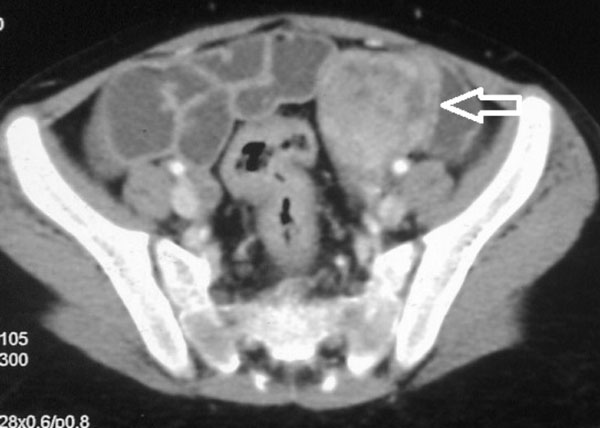
raised (19 ng/mL/h; normal 0.5-6 ng/mL/h). Computed tomography (CT)
abdomen with CT angiography revealed normal bilateral kidneys and renal
arteries, but incidentally detected a well circumscribed lobulated soft
tissue mass (4.1 × 3.4 cm) in left adnexa showing heterogeneous poor
contrast enhancement (Fig. 1). A 24-hour urinary
catecholamines report was normal; vanillylmandelic acid 3.85 µcg (normal
0-15), adrenaline 3.5 µcg (normal 0-20) and nor-adrenaline 26.25 µcg
(normal 0-90). Serum catecholamine measurement showed elevated
nor-adrenaline (916.66 pg/mL; normal 0-600 pg/mL) and normal adrenaline
(58.3 pg/mL; (normal 0-100 pg/mL). Based on CT finding and serum
catecholamine report, diagnosis of left ovarian paraganglioma was made.
123Metaiodobenzyl-guanidine
scan did not show any increased tracer uptake.
 |
|
Fig. 1 CECT abdomen showing a well
circumscribed 4.1 × 3.4 cm lobulated soft tissue mass in left
adnexa (arrow) showing heterogeneous poor contrast enhancement.
|
As the child presented with hypertensive emergency,
injection labetalol was administered, which was later substituted with
oral formulation. Subsequently, other antihypertensives were added in
combination (Amlodipine, Clonidine and Enalapril). Despite using
multiple antihypertensives, BP remained elevated above 95th centile.
Prazosin was added in the pre-operative period, which resulted in near
normalization of BP.
The child underwent open laparotomy and left
salpingo-oophorectomy was performed. An ovarian mass measuring 5x4x3.5
cubic centimeter with an external pearly white capsule was removed.
Microscopic examination revealed a monomorphic population of round cells
with abundant granular cytoplasm arranged in the classical zellballen
pattern. On immunohisto-chemistry, tumor cells expressed synpatophysin
and cytokeratin (Web Fig. 1). A final diagnosis of
ovarian paraganglioma was made based on histopathology report. Two weeks
after surgery, her BP was normal (112/66 mm Hg), polyuria subsided and
biochemical parameters (serum Na and K) normalized. Antihypertensives
were gradually tapered and omitted over next 6 weeks. At last follow-up
(2 years after surgery), she was normotensive, off medication and had
not shown any sign of tumor recurrence.
Discussion
Paragangliomas are rare extra-adrenal
catecholamine-secreting tumors located in paravertebral axis and
sympathetic nerve branches in pelvic organs, and secrete
nor-epinephrine. Parasympathetic variety is located in head and neck,
and are generally non-secretory [5]. Paragangliomas have been rarely
reported in ovary, uterus and vagina, constituting only 2% of all
gynecological tract tumors [6-8]. Clinical features are often
nonspecific and tumor detection may be incidental. Polyuria and
polydipsia as presenting complaints have been reported earlier in a
9-year-old boy with pheochromocytoma [9].
The child in our report presented with polyuria,
polydipsia, hypertension and hyponatremia – similar to clinical and
biochemical features of HHS. Release of natriuretic peptides (BNP and
ANP) with rapid elevation of BP might have resulted in polyuria and
hyponatremia by pressure natriuresis. Natriuretic peptides suppress
sodium reabsorption at thiazide-sensitive distal convoluted tubules and
thereby increase its delivery to the downstream collecting ducts, where
aldosterone stimulates secretion of potassium resulting in hypokalemia
[10]. Unlike renovascular hypertension, where renal ischemia is central
to activation of renin-angiotensin system (RAS) and subsequent HHS, in
our case, hypovolemia secondary to polyuria might have resulted in
activation of RAS. We postulate that intense thirst and release of
anti-diuretic hormone due to hypovolemia resulting from polyuria could
have also contributed to the development of hyponatremia in our case.
Proteinuria could have resulted from glomerular hyperfilteration
secondary to hypertension; resolution of proteinuria with BP control
after surgery validates the same.
Paragangliomas can occur sporadically or in
association with familial syndromes like multiple endocrine neoplasia
(MEN) type 2, Von Hippel-Lindau disease, and neurofibromatosis type 1.
Our case did not have any features suggestive of familial syndromes.
However, genetic testing, urinary/plasma metanephrines, plasma
aldosterone and FDG PET could not be performed because of financial
constraints.
Treatment of paragangliomas is chiefly surgical.
Adequate preoperative BP control is mandatory to avoid intra operative
rise in BP due to excessive release of catecholamines. Use of selective
alpha-1 adrenergic receptor blocker in preoperative period is more
rationale and effective way to control BP in children with
pheochromocytoma/paraganglioma. Beta blockade is instituted following
alpha blockade to offset reflex tachycardia and should never be started
before adequate alpha blockade to prevent risk of severe hypertensive
crisis from unopposed alpha-1 receptor stimulation [5].
To conclude, HHS may be a manifestation of
paraneoplastic syndrome. A high index of clinical suspicion, CT or MRI
of abdomen, and estimation of urinary and serum catecholamines may help
to clinch these rare causes.
Contributors: MK,AD,KM: initial work-up and case
management; VM: performed the surgery; NM: made the histopathological
diagnosis; AD: prepared the initial draft; MK,NM,KM: revised the draft.
All the authors approved the final version of the manuscript.
Funding: None; Competing Interest:
None stated.
References
1. Tullus K, Brennan E, Hamilton G, Lord R, McLaren
CA, Marks SD, et al. Renovascular hypertension in children.
Lancet. 2008;371:1453-63.
2. Pandey M, Sharma R, Kanwal SK, Chhapola V, Awasthy
N, Mathur A, et al. Hyponatremic-hypertensive syndrome: Think of
unilateral renal artery stenosis. Indian J Pediatr. 2013; 80:872-4.
3. Mukherjee D, Sinha R, Akhtar MS, Saha AS.
Hyponatremic hypertensive syndrome – a retrospective cohort study. World
J Nephrol. 2017;6:41-44.
4. Kovalski Y, Cleper R, Krause I, Dekel B, Belenky
A, Davidovits M. Hyponatremic hypertensive syndrome in pediatric
patients: is it really so rare? Pediatr Nephrol. 2012;27:1037-40.
5. Bholah R, Bunchman TE. Review of pediatric
pheochromocytoma and paraganglioma. Front Pediatr. 2017; 5:155.
6. Kefell M, Usubütün A. An update of neuroendocrine
tumors of the female reproductive system. Turk J Pathol. 2015;31:128-44.
7. Liu H, Li WZ, Wang XY, Pei YG, Long XY, Chen CY,
et al. A rare case of extra-adrenal pheochromocytoma localized to
the ovary and detected via abdominal computed tomography angiography.
Oncol Lett. 2015;9:774-6.
8. Mc Cluggage WG, Young RH. Paraganglioma of the
ovary: report of three cases of a rare ovarian neoplasm, including two
exhibiting inhibin positivity. Am J Surg Pathol. 2006;30:600-5.
9. Jain V, Yadav J, Satapathy AK. Pheochromocytoma
presenting as diabetes insipidus. Indian Pediatr. 2013;50:1056-7.
10. Nagakawa H, Mizuno Y, Harada E, Morikawa Y, Kuwahara K, Saito Y,
et al. Brain natriuretic peptide counteracting the
Renin-angiotensin-aldosterone system in accelerated malignant
hypertension. Am J Med Sci. 2016;352:534-39.
|
|
|
 |
|
