A 10-year-old boy presented with a solitary pruritic skin
lesion near the right eye which was present since 4 months.
To start with, there was a small elevated lesion with mild
itching which gradually increased in number and coalesced.
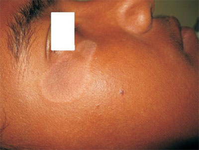
Cutaneous examination revealed a well circumscribed 3cm X
1.5cm plaque with lichenification towards the centre. The
margin of the lesion was hypopigmented whereas the centre
was hyperpigmented. Fine scales were noted in the periphery
of the lesion (Fig. 1). No similar lesion was
present elsewhere in the body; hairs, nails and mucosae were
normal. Biopsy showed epidermal spongiosis with a dermal,
perivascular, mainly mononuclear cell infiltrate and edema.
A diagnosis of plaque-type polymorphous light eruption was
made.
 |
|
Fig. 1 Solitary
well-circumscribed plaque on face.
|
Polymorphous light eruption is an
acquired sunlight-induced dermatosis, particularly at
temperate latitudes, affecting 10-20% of the population. It
is usually characterized by an itchy, erythematous,
symmetrically distributed, papulovesicular rash, on some
exposed areas within hours of exposure to ultraviolet
radiation. Classical histopathological findings include
epidermal spongiosis with a dermal, perivascular, mainly
mononuclear cell infiltrate and edema. It usually responds
to broad-spectrum sunscreens and oral or topical steroids.
Prophylactic low-dose immunosuppressive phototherapy in
spring may be given for frequent episodes. Close clinical
differentials are Hansen’s disease (presence of hypoesthesia
or anesthesia) and psoriasis (silvery white scales over an
erythematous plaque).


