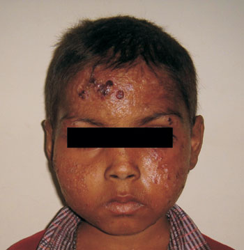
A 5 year old boy presented with papules and nodules on face, chest and upper extremities and seborrhea of scalp for 4 years. Some old healed scars were also present. Lesions were pruritic to begin with but were non-itchy. Examination revealed erythematous plaques and nodules on face and upper limbs. Lesions were more marked on forehead, cheek (Fig. 1) and dorsum of hands. There were contractures of little fingers of the both hands (Fig. 2). Lymphadenopathy and hepatosplenomegaly were absent. Histopathological examination from skin lesions was suggestive of langerhans cell histiocytosis (LCH). Immunohistochemical studies showed positive staining with antibodies to S-100 and CD1a, consistent with the diagnosis of LCH. A complete bone survey, CT cranium and chest, ultrasonography abdomen and bone marrow examination were negative for systemic involvement. The final diagnosis was cutaneous LCH without systemic involvement. After a trial of topical steroids which were ineffective, he was started on oral methotrexate 20 mg/m2 weekly for 6 months. Child had marked improvement with complete resolution of the lesions within 6 weeks. He is in regular follow up and there is no recurrence of lesions.
 |
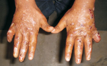 |
Fig.1 Erythematous plaques and nodules on face. |
Fig.2 Papules and nodules on dorsum of hand. |
Langerhans cell histiocytosis is a clonal proliferative disorder of langerhans cells typically seen in infants and children. The manifestations are protean and can involve virtually every tissue in the body. The clinical presentation of cutaneous LCH is variable. It can present as papules, plaques or nodules localized to a single anatomic site or in a generalized distribution. Systemic involvement of bones, lungs, liver, lymph nodes, hypothalamic pituitary axis or central nervous system can occur several years following initial skin lesions. The differential diagnosis includes seborrheic dermatitis, psoriasis, lupus miliaris faciei, granuloma faciale and xanthoma disseminatum. Biopsy from skin lesions is essential for definitive diagnosis. Treatment options range from observation alone to topical steroids and oral therapy with methotrexate and thalidomide. Psoralen and long-wave ultraviolet radiation have also been tried. Patients with limited cutaneous involvement have a better prognosis than those with systemic disease and prognosis correlates with the extent of systemic disease.
|


