Malignant Otitis externa (MOE) is a life threatening, progressive
bacterial infection of the external auditory canal (EAC), mastoid and
skull base. It most commonly occurs in elderly diabetics or in an
otherwise immune compromised host. In nearly all cases Pseudomonas
aerugenosia is the causative organism(1,2). We report a case of
malignant otitis externa caused by Enterobacter in a 10 month old
immunocompetent infant.
A 10 month old girl, second sibling of non
consanguineous parents, presented with complaint of purulent ear discharge
from left ear since 2 months, intermittent fever of 100 – 101ºF and
deformation of left ear since 1 month. There was no history of trauma or
ear picking. Patient had already received 10 days of parenteral and local
antibiotics with no response.
The girl was well nourished, had mild pallor and was
febrile and irritable. The left ear was deformed with necrotic material
seen all over the left external ear. On examination granulation tissue was
seen occluding the external ear with erythema surrounding the pinna (Fig.1).
Tympanic membrane could not be seen. There was no abscess collection on
the surrounding areas and mastoid tenderness was not present. There was no
cranial nerve palsy or other intracranial complication. CT scan of left
temporal bone showed destruction of complete cartilageous and some bony
part of external auditory canal with secondary opacification of mastoid
air cells. Right side was normal.
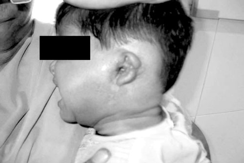 |
|
Fig.1
Malignant Otitis Externa: Markedly
deformed left ear with granulation and purulent tissue occluding the
external ear. |
Pus culture from ear swab revealed Enterobacter
species after which antibiotics were modified according to sensitivity
pattern. Local treatment with normal saline compresses were given. Fever
and erythema subsided within 1 week. There was narrowing of the opening of
the external auditory canal for which stent was placed to prevent complete
closure. Patient was discharged after 3 weeks of parenteral antibiotics on
oral antibiotics for 3 more weeks. Reconstruction of the deformed external
ear was planned at a later date.
The diagnosis of malignant otitis externa is by two
criteria: obligatory and occasional. The obligatory criteria are pain,
edema, exudates, granulations, micro abscess (when operated), positive
bone scan or failure of local treatment for more that 1 week, and
possibility of Pseudomonas in culture. The occasional criteria
include diabetes, cranial nerve involvement, positive radiograph,
debilitating condition and old age(3). Our patient fulfilled the
obligatory criteria.
Although rare, malignant otitis externa has been
reported in children with diabetes and other immune compromised
states(4,5). Complications include necrosis of the tympanic membrane,
stenosis of EAC, auricular deformity and sensorineural and conductive
hearing loss. Isolation of Enterobacter has not been reported
earlier. Prolonged treatment with sensitive antibiotics is recommended for
6 to 8 weeks. Inadequate treatment can lead to recurrence of disease.
Quinolones are generally avoided. Treatment can be guided by monitoring
ESR and Gallium scans(6).
Acknowledgment
We are thankful to Dr Daljit Singh, Principal and
Professor (Pediatrics), for his guidance.
References
1. Meltzer PE, Kelemen G. Pyocyaneous osteomyelitis of
the temporal bone, mandible and zygoma. Laryngoscope 1959; 69: 1300-1316.
2. Midwinter KI, Gill KS, Spencer JA, Fraser ID.
Osteomyelitis of the temporomandibular joint in patients with malignant
otitis externa. J Laryngol Otol 1999; 113: 451-453.
3. Cohen D, Friedman P. The diagnostic criteria of
malignant external otitis. Laryngol Otol 1987; 101: 216-221.
4. Paul AC, Justus A, Balraj A, Job A, Kirubakaran CP.
Malignant otitis externa in an infant with selective IgA deficiency: a
case report. Int J Pediatr Otorhinolaryngol 2001; 60: 141-145.
5. Sobie S, Brodsky L, Stanievich JF. Necrotizing
external otitis in children: report of two cases and review of the
literature. Laryngoscope 1987; 97: 598-601.
6. Carfrae MJ, Kesser BW. Malignant otitis externa. Otolaryngol Clin
North Am 2008; 41: 537-549.


