|
The management of a
neonate with a known difficult airway is a
challenge to any anaesthesiologist. There is
always the fear of loss of control over the
airway and difficult reintubation following
rigid bronchoscopy. Preparation, therefore,
should include a plan for safe reintubation. We
report a case of a four-day-old neonate who
presented to us for rigid bronchoscopy and an
innovative technique to secure the airway and
reintubate the child.
Case History
A four-day-old neonate
presented to our hospital for evaluation of the
airway using a rigid bronchoscope. The neonate
was full-term and delivered by cesarean section
for fetal distress. He had poor respiratory
effort and bradycardia at birth and required
endotracheal intubation. The initial attempts
for endotracheal intubation by a pediatric
resident failed. The airway was finally secured
with a 2.5 endotracheal tube (ETT) after
multiple traumatic attempts by a senior
pediatrician. The neonate was ventilated and
shifted to our center on the fourth day for
evaluation of airway congenital anomalies. The
systemic examination was normal and the neonate
was breathing spontaneously and moving all
limbs. The chest X-ray confirmed the
position of the endotracheal tube and no
abnormality was noted. The pediatrician who
intubated the child was contacted and he opined
that it was an extremely difficult airway to
access as there was probably an anatomical
distortion and blood in the upper airway. The
prenatal ultrasound scans did not indicate any
anomalies. The risk of losing control of the
airway was discussed with the surgeons and a
backup plan tracheostomy was made, if
reintubation was difficult.
On arrival to the theatre and
under routine monitoring, the child was induced
with sevoflurane 1-6% in 100% oxygen. A leak
test was performed and an audible air leak was
found around the ETT at pressure of less than 15
cm H2O.
Direct laryngoscopy with a miller 1 blade showed
intraoral edema and only the tip of the
epiglottis could be visualized. Anticipating a
difficult airway, we did not paralyse the child
and introduced a central venous line (CVP)
guidewire (60 cm and 0.89 mm) into the ETT to
the length of approximately 15 cm. Extubation
was done over the guide wire and a size 3
ventilating rigid bronchoscope was introduced
into the trachea by the side of the guide wire.
On bronchoscopy, no congenital anomalies were
revealed, except for glottic edema and the
guidewire had not caused any injuries. At the
end of the procedure, direct laryngoscopy was
done and the larynx was visualized with a Lehane
and Cormack grade 3 view and the trachea was
reintubated easily with a 3.5 ETT railroaded
over the guidewire. ETT placement was confirmed
by auscultation and capnography. A leak test was
performed and an audible leak was present at
pressure of 20 cm H2O.
The neonate was shifted to the Neonatal
Intensive Care unit after the return of
spontaneous breaths and ventilated
postoperatively. The neonate was nebulised with
adrenaline and parenteral steroids were
administered post procedure. Post-operative
chest X-ray had no radiological
abnormalities. He had an uneventful recovery and
was extubated after two days.
Discussion
Rigid bronchoscopy is a safe
technique in the neonatal/infant period for the
diagnosis of airway pathology and it directs
early management of these cases [1]. It gives an
excellent view of the large airways, but
requires general anaesthesia/sedation and the
neonate has to be extubated to introduce the
bronchoscope. Reintubation may be a challenge in
neonates with a known difficult airway and may
end up as a catastrophe if there is a delay in
securing the airway.
Reintubation at the end of
the procedure by conventional methods in this
neonate was anticipated to be difficult because
of severe intraoral edema caused by multiple and
prolonged airway instrumentation, also due to
the suspicion of congenital anomalies. In
addition the neonate had not received any
steroid to reduce airway edema at the referring
hospital. Dexamethasone in a dose of 0.25-0.5
mg/kg iv; 3-5 doses starting at least 6-12 hours
is indicated for elective extubation of "high
risk" neonates, before extubation [2]. We have
used a guidewire to prevent the loss of airway,
to serve as a guide for rigid bronchoscopy and
to facilitate endotracheal intubation at the end
of procedure without interfering with the rigid
bronchoscopy.
Guidewires have been safely
used in many scenarios for facilitating
endotracheal intubation. The CVP guidewire has
been used as a guide for changing an oral tube
to a nasal tube in the absence of sophisticated
equipment [3]. They have also been used for
retrograde intubation in infants as young as one
month [4]. Scherlitz and Peters [5] have
reported two cases with difficult airway, where
a guidewire was left in the trachea
postoperatively for assisting in rapid
reintubation in the immediate postoperative
period, if required. Guidewires have been
introduced anterograde through the Laryngeal
Mask Airway for subsequent endotracheal
intubation [6,7]. Rodriguez, et al. [8]
used a guidewire through the working channel of
the fiberoptic bronchoscope (FOB) for
railroading an ETT in a case of Treacher Collins
syndrome.
 |
|
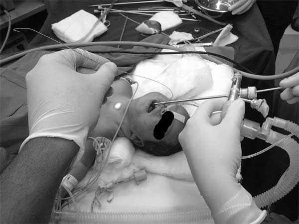
Fig. 1 The
guide wire held in place by the
anaesthesiologist. Rigid bronchoscopy
being done by the surgeon;
transillumination can clearly be seen in
the anterior aspect of the neck.
|
The guidewire is long (60
cm), smooth, and sufficiently rigid to allow
"railroading" of tracheal tube over it. The
small outer diameter of the guidewire allows a
tracheal tube as small as 2.5 mm. A rigid
bronchoscope can easily be introduced into the
trachea by the side of the guidewire. The
guidewire is visible throughout the procedure
and the anaesthesiologist has control over its
movement or dislodgement. The main disadvantage
of this technique is inability to provide
supplemental oxygen if there is a delay in
reintubation and although it is a blunt wire
there is always a small risk of lower airway
injuries. We have used the straight end of the
guidewire rather than the J tip end as we
anticipated that the J tip may get entangled
with the rigid bronchoscope or may get entrapped
in the bronchi during bronchoscopy. Though there
is a small possibility of CVP guidewire related
complications (kinking, knotting or
perforation), we had to use it as our options
were limited. A pediatric FOB or a neonatal
airway exchange catheter (AEC) was not available
in our institution.
The use of ultra-thin
pediatric FOB for assessment of tracheo-bronchial
injuries and congenital abnormalities is a safer
alternative to rigid bronchoscopy [9-10]. In a
developing country like ours, very few tertiary
care centers have an ultra thin FOB. The use of
AEC airway exchange catheters (AEC) for
difficult extubation is safe technique, but was
not feasible in this scenario as the AEC would
interfere with space required for introduction
of the bronchoscope.
In conclusion, the use of a
CVP guidewire to prevent the loss of airway and
to reintubate neonates/infants at the end of
rigid bronchoscopy is an innovative, safe, cost
effective and successful technique.
Contributors: All the
authors have contributed, designed and approved
the manuscript. VSP: will act as guarantor.
Funding: None;
Competing interests: None stated.
References
1. Prinja N, Manoukian JJ.
Neonatal/infant rigid bronchoscopy. J
Otolaryngol. 1998;27:31-6.
2. Markovitz BP, Randolph AG,
Khemani RG. Corticosteroids for the prevention
and treatment of post-extubation stridor in
neonates, children and adults. Cochrane Database
Syst Rev. 2008;16:CD001000.
3. Sharma R, Kumar A, Panda
A. Using a central venous pressure guidewire and
suction catheter to facilitate oral to nasal
tracheal tube change in a child with a difficult
airway. Anesth Analg. 2009;108:1716–7.
4. Schwartz D, Singh J.
Retrograde wire-guided direct laryngoscopy in a
1-month-old infant. Anesthesiology. 1992;77:607.
5. Scherlitz A, Peters J. A
guidewire as a reintubation aid. Translaryngeal
fiberoptic insertion of a guidewire into the
trachea to assist fiberoptic reintubation in
patients difficult to intubate. Anaesthetist.
1994;43:618-20.
6. Nitahara K, Watanabe R,
Katori K, Yamasato M, Matsunaga M, Dan K.
Intubation of a child with a difficult airway
using a laryngeal mask airway and a guidewire
and jet stylet. Anesthesiology. 1999;91:330-1.
7. John B, Linga-Nathan P,
Mendonca C. Tracheal intubation through a single
use laryngeal mask airway using a guidewire
technique. Can J Anaesth. 2007;54:775-6.
8. Rodriguez AM, Etxaniz A,
Rey AM, Perez J, Nieto CM. Use of a metal guide
in the working channel of a fiberoptic scope to
insert a tracheal tube in an infant with
Treacher Collins syndrome and choanal atresia.
Rev Esp Anestesiol Reanim. 2010;57:115-8.
9. Schelhase DE, Graham LM,
Fix EJ, Sparks LM, Fan LL. Diagnosis of tracheal
injury in mechanically ventilated premature
infants by flexible bronchoscopy. Chest. 1990;
98:1219-25.
10. de Blic J, Delacourt C,
Scheinmann P. Ultrathin flexible bronchoscopy in
neonatal intensive care units. Arch Dis Child. 1991;66:1383-5.
|


