|
|
|
Indian Pediatr 2014;51: 677-678 |
 |
Acute Hemorrhagic Edema of Infancy
|
|
Abhijit Dutta And *Sudip Kumar Ghosh
Department of Pediatric
Medicine; North Bengal Medical College; and
*Department of Dermatology, Venereology and Leprosy,
RG Kar Medical College,West Bengal, India.
Email:
[email protected]
|
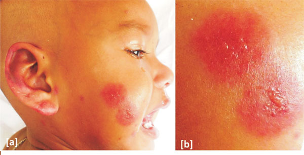
A 14-month-old girl presented with acute onset erythematous
skin eruption on her body, following an episode of upper
respiratory tract infection. On examination, the child was
febrile and the vitals were stable. There were multiple,
non-tender, purpuric targetoid lesions studded with vesicles
on her face, pinna, extremities and buttocks. The mucosa and
the trunk were spared. There was mild non-pitting edema over
the upper extremities and the face. Systemic examination was
normal. Routine blood examination, coagulation profile,
renal function tests, blood culture, and urine analysis were
normal, except for mild leucocytosis (total leukocyte count
12,600 mm
3).
Histopathlogical examination from the lesion showed features
of leucocytoclastic vasculitis. A diagnosis of Acute
hemorrhagic oedema of infancy (AHEI) was made. Fever
subsided in two days and the skin lesions completely
subsided within the next two weeks.
 |
|
Fig. 1 (a) Purpuric
targetoid lesions on face; (b) Close-up view of the
lesion.
|
AHEI is a benign, self-limiting
leucocytoclastic vasculitis generally affecting children
under the age of 2 years. An upper repiratory illness
usually precedes the sudden onset of red macules or
urticarial skin lesions. AHEI lesions vary from 0.5 to 4 cm
in size occasionally becoming confluent to annular or
targetoid purpuric lesions. It mainly affects the face and
extremities, sparing the trunk, and often accompanied by
non-pitting edema.
Differential diagnosis of AHEI include
Henoch Schonlein purpura (older age, smaller lesions, facial
sparing, systemic involvement, slow resolution,
meningococcemia (central necrosis), erythema multiform
(three concentric color zones), Sweet’s syndrome (erythematous
blue or violet papules, plaques, or nodules often with a
pseudo-vesicular appearance), urticarial vasculitis (absence
of target-like lesions; purpuric spots visible on diascopy,
hyperpigmentation on healing), and fixed drug eruptions
(round or oval sharply delineated erythematous plaques with
central blister or necrotic area).
|
|
|
 |
|
