|
|
|
Indian Pediatr 2014;51:
316-317 |
 |
Pemphigus Vulgaris in a Neonate and his Mother
|
|
Sheethal S Kodagali, SD Subbarao and R Hiremagaloor
From the Department of Pediatrics, Dr Malathi Manipal
Hospital, Bengaluru, India.
Correspondence to: Dr Sheethal S Kodagali, Department
of Pediatrics, Dr. Malathi Manipal Hospital,
# 45/1, 45th cross, 9th block, Jayanagar, Bangalore - 560 069, India.
Email: [email protected]
Received: July 10, 2013;
Initial review: August 5, 2013;
Accepted: March 10, 2014.
|
|
Background: Neonatal pemphigus is a rare, transient blistering
condition due to transplacental transfer of maternal autoantibodies.
Case characteristics: A male neonate born to a mother with oral
pemphigus was noticed to have multiple lesions. Observation:
Multiple flaccid bullae were noticed on the face, scalp, trunk and
extremities with clear fluid and few areas of erosions. Outcome:
All lesions resolved at the end of one week with conservative
management. Message: Maternal pamphigus may rarely involve her
newborn infant; it resolves on its own.
Keywords: Autoimmunity, Neonatal pemphigus
vulgaris, Transplacental transfer.
|
|
P
emphigus is a group of
autoimmune blistering disease of skin and mucous membranes [1].
Incidence rates between 0.1 and 0.5 per 100,000 people per year have
been reported [2]. Neonatal pemphigus is a transient autoimmune
blistering disease caused by transfer of maternal IgG autoantibodies to
desmoglein-3 through the placenta when the mother is affected with
pemphigus [3,4].
Case Report
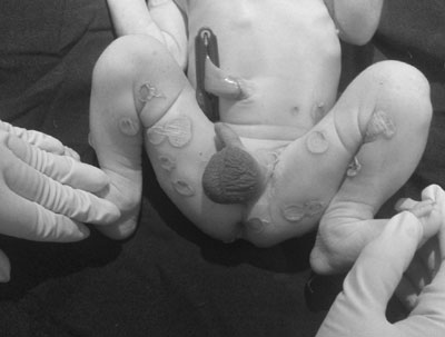
A 1-day-old boy weighing 2900 grams, born after full
term pregnancy, was noticed to have multiple flaccid bullae on the face,
scalp, trunk and extremities with clear fluid and few areas of erosions.
All lesions showed a rim of erythema and abrupt demarcation from the
surrounding normal skin (Fig. 1). These lesions were not
restricted to trauma-prone areas. A few lesions had profuse serous
discharge. Nails and oral mucosa were not involved. Mother of this
infant was diagnosed to have oral pemphigus vulgaris 8 months before
conception, documented by incisional biopsy from buccal mucosa and
direct immunofluorescence test (DIF), and was on daily oral steroids.
The child was suspected to have neonatal pemphigus vulgaris based on the
morphology and distribution of skin lesions, and maternal history. A
differential diagnosis of herpes simplex, candidosis, syphilis,
infectious mononucleosis and epidermolysis bullosa were also considered.
Tzanck smear was negative for multinucleated giant cells and
acantholytic cells. Mother’s VDRL test was negative. Skin biopsy was
deferred as the lesions were drying up on second day. Child was managed
with warm saline compresses, barrier nursing, topical antibiotics and
breast feeds supplemented with formula feeds. Intravenous fluids were
not required. Fluid input, output and electrolytes were monitored
regularly. All the lesions resolved at the end of one week.
 |
|
Fig.1 Flaccid bullae and crusted
erosions with an erythematous rim distributed over the groin
area in the neonate.
|
Discussion
Pemphigus is defined as a group of life-threatening
blistering disorders characterised by acantholysis (loss of keratinocyte
to keratinocyte adhesion) that results in the formation of
intraepithelial blisters in mucous membranes and skin. The process of
acantholysis is induced by circulating autoantibodies to intracellular
adhesion molecules [2]. Patients with pemphigus develop mucosal erosions
and/or flaccid bullae, erosions, or pustules on skin. Neonatal pemphigus
is a very rare transient form which occurs as a consequence of placental
transmission of autoantibodies to the fetus from the mother. Maternal
Pemphigus causes premature births and still births, with rare occurrence
of neonatal pemphigus. The prognosis is very good with resolution of
lesions completely by 3 weeks of life [5].
References
1. Jacqueline P, Scott RF, Jason H, John Z, Kristin
L, Shery V. Neonatal pemphigus in an infant born to a mother with
serologic evidence of both pemphigus vulgaris and gestational pemphigoid.
J Am Acad Dermatol. 2009;60:1057-62.
2. Michael H, Cassian S. Pathogenesis, Clinical
Manifestations and Diagnosis of Pemphigus. Available from: URL: http//
www.uptodate.com/store. Accessed July 7, 2013.
3. Sameera BKI, Yashodhara BM, Shashikiran U.
Pemphigus vulgaris in a pregnant woman and her neonate. BMJ.
2012:2013:1-5.
4. Moncada B, Kettelsen S, Hernandez JL, Ramirez F.
Neonatal pemphigus vulgaris: role of passively transferred pemphigus
antibodies. Br J Dermatol. 1982; 106:465-8.
5. Ian AG. Maternal Medicine: Medical Problems in
Pregnancy, Dermatological disease in pregnancy, 1st ed. UK: Elsevier
Health Sciences. 2007. P.276.
|
|
|
 |
|
