A 3-years-old female child presented with asymptomatic multiple
well-defined erythematous scaly plaques since infancy. She was born of a
non-consanguineous marriage and had uneventful prenatal and natal period.
The lesions started appearing in first few months of life first on knees
and then over rest of the body. The lesions were persisting in nature and
she was never lesion free. However, the appearance (erythema and
thickness) used to improve at times, only to get worse soon after. Rest of
the history was non-contributory and no other family had similar lesions.
There was no history of appearance of transient erythematous lesions. On
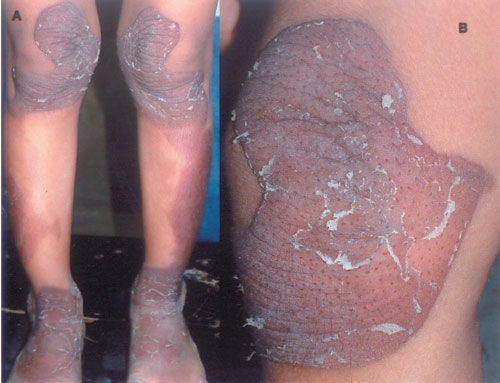
examination, erythematous scaly plaques were present on extensor aspect of
extremities (knees, lateral leg, ankle, and elbows) and sacral region with
striking symmetry (Fig. 1).
 |
|
Fig. 1 (a) Well demarcated
symmetrical erythematous plaques over lower extremities. (b) Close
up of a lesion. |
Face, trunk, palm, sole, mucosa, hair, and nails were
lesion free. Differential diagnoses included psoriasis, pityriasis rubra
pilaris (PRP) (circumscribed type), erythrokeratodermia variabilis (EKV),
and progressive symmetric erythrokerato-dermia (PSEK). Clinically,
psoriasis (absence of significant scaling and negative Auspitz sign), PRP
(absence of any follicular keratotic lesions) and EKV (no history of
transient erythematous lesions) were ruled out. Histopathology findings
were consistent with the diagnosis of PSEK. This condition is
characterized by erythematous plaques that appear shortly after birth,
progress slowly during the first few years, and then stabilize in early
childhood. The transient migratory erythema that defines EKV is absent. It
is transmitted in autosomal dominant manner and mutation in protein
loricrin (envelope protein) has been found in one family. There is no
specific treatment, though emollients and keratolytics provide cosmetic
improvement.


