|
|
Case Reports Indian Pediatrics 2006;43:908-910 |
||
|
Symptomatic Hepatic Hemangioendothelioma in a Newborn |
||
|
K. Jothilakshmi From the Department of Pediatrics & *Pediatric Surgery, P.S.G. Institute of Medical Sciences & Research, Peelamedu, Coimbatore 641 004, Tamil Nadu, India.
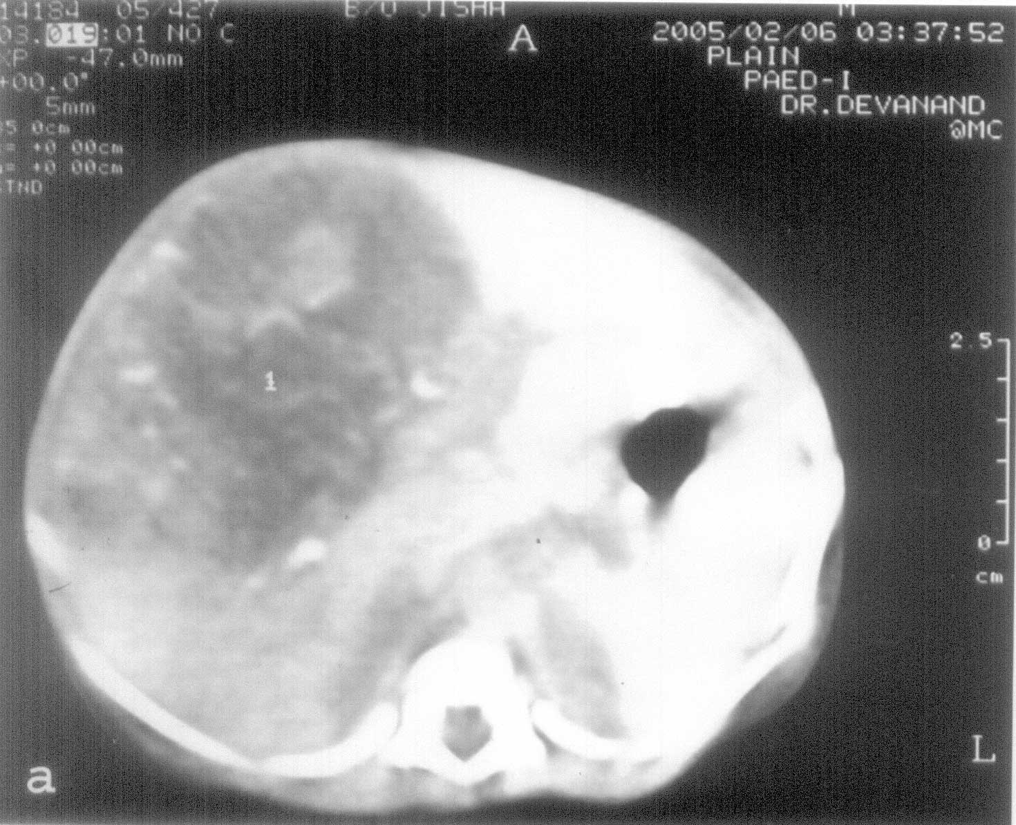
Benign infantile hemangioendothelioma is a rare neonatal hepatic tumor. They may undergo spontaneous regression, but can sometimes be life threatening due to congestive cardiac failure and/or consumptive thrombocytopenia. While in asymptomatic cases observation is the recommended course, for patients with symptoms optimum management strategy is still controversial. We report a sick neonate with hepatic hemangioendothelioma who responded to high dose steroids. Case Report A 2.3 kg female neonate delivered by LSCS at 36 weeks for oligohydramnios and premature rupture of membranes was referred for a visible lump in the right hypochondrium at birth. The mother’s antenatal period was uneventful and she had no antenatal sonography. At 4 hours, the baby was lethargic, had respiratory distress, capillary refill time was more than 3 seconds and peripheral pulses were weak. The heart rate was 170/min. There was no gallop rhythm or murmur. A 5 × 5 cm lump was seen in the right hypochondrium which was firm, non tender and non-pulsatile. The liver was enlarged almost up to the right iliac fossa and spleen was just palpable. There was no bleeding manifestations or cutaneous hemangiomas. She had recurrent subtle seizures in addition to brief multi focal clonic seizures. The blood sugar was 16 mg/dL. Serum calcium, urea, creatinine and electrolytes were normal. Her hematological profile was unremarkable except for a platelet count of 88,000/mm3. Chest X-ray revealed a cardio-thoracic ratio of 60% with clear lung fields. An ECHO cardiogram showed no cardiac anomaly. The abdominal sonography (USG) showed a lobulated heterogeneously enhancing mass in the liver with increased vascularity involving segments 4-8. Contrast enhanced CT scan of the abdomen revealed a heterogenosly hypodense mass with dense peripheral enhancement after contrast injection while the central area remained hypodense. There were area of scattered calcification also. (Fig.1). The blood a-feto protein (AFP) was 9OO IU/mL. The serum bilirubin was 18.4 mg/dL (direct 12 mg/dL).
There is no recurrence of seizure after correcting the hypoglycemia. The cardiac failure was managed with fluid restriction, oxygen, diuretics and ionotropic support. There was a progressive drop in platelet count to 44,000/cu mm over the next 3 days. Oral prednisolone was started in a dose of 4 mg/kg/day in 2 divided doses. There was improvement in general physical condition and platelet count stopped declining and normalized by day 10. The bilirubin level decreased but, conjugated hyperbilirubinemia persisted even after control of congestive cardiac failure. On day 7, ursodeoxycholic acid and phenobarbitone was started as cholerectic agents. Serum bilirubin normalized by day 14. Prednisolone was tapered to 4 mg/kg/day on alternate days after 2 weeks. It was further tapered to 2 mg/kg on alternate days by 6 weeks and 1 mg/kg on alternate days at 3 months. At 3 months follow-up, the liver was just palpable and the USG showed a small 3 cm mass with some vascularity and baby was thriving well. Discussion Liver tumors are rare and account for 5% of all neoplasms in the fetus and newborn. Infantile hemangio-endotheliomas, cavernous hemangiomas, mesenchymal hematomas and hepatoblastomas are the most frequent. Majority of the neonatal liver tumors are benign vascular neoplasms. Most of these are incidentally detected in USG. However, infantile hemangioendotheliomas in particular can cause severe symptoms such as abdominal distension, gross hepatomegaly, severe AV shunting with congestive cardiac failure, anemia, thrombocytopenia (Kasabach-Merrit syndrome), consumptive coagulopathy, intra- abdominal hemorrhage, etc. Rarely, biliary obstruction with jaundice(1,2), vomiting and gastric outlet obstruction(3) have been reported. Initial investigation for diagnosis is USG with Doppler evaluation. It may show a single or more commonly, multiple hyper-echoic lesions scattered in the liver with increased vascularity. There may be an enlarged celiac axis with abrupt tapering of the aorta distal to it. This is considered highly suggestive of hemangioendothelioma. A more definite diagnosis requires either a contrast enhanced CT scan or MRI. Classical picture of contrast enhanced CT (CECT) is a hypodense area which enhances on contrast injection, though some lesions with a central area of hemorrhage or necrosis may not show enhancement. MRI picture resembles CECT scan and avoids the use of contrast and radiation. Selective angiography of the celiac and hepatic artery delineates the vascular supply and is useful when embolisation is contemplated. Serum AFP should be done in all cases, though the levels in infants are above normal till 6 months of age. AFP levels may be high in neonates with hemangioendothelioma but they are never as high as seen in hepatoblastoma. In case a malignant liver mass cannot be excluded beyond doubt, it may be reasonable to proceed with open biopsy of the mass. The management of hemangioendotheliomas is
controversial. Asymptomatic lesions can be followed up, using serial USG
to visualize the anticipated spontaneous regression. Medical treatment
includes control of CCF, administration of blood products to correct
anemia and coagulopathy. Steroids (prednisolone) 2-5 mg/kg/day, can be
tried to suppress continued growth of the lesion and encourage
regression after the desired effect is achieved. They can be continued
for 2-3 weeks and tapered slowly over 2 -3 months. Systemic
corticosteroids have become a mainstay in the treatment of
hemangio-endotheliomas. Yet, their mechanism of action is not well
understood(4-8). Daily doses of 2-3 mg/kg of prednisolone are usually
given and some investigations have recommended higher doses of 5
mg/kg/day(9). This treatment results in dramatic shrinkage of the
hemangioma in one third or a third may show no responsive lesions
unresponsive to steroids can be treated with a-2a interferon.
Invasive measures like hepatic artery ligation or embolisation may be
tried with rapid onset of severe symptoms. Small solitary tumors may be
best treated by complete resection. | ||
|
References | ||
|
![]()