|
|
|
Indian Pediatr 2016;53: 1037 |
 |
Retinoblastoma Mimicking Orbital Cellulitis
|
|
Anirban Das, #Usha
Singh and *Deepak Bansal
Pediatric Hematology-Oncology unit, Department of
Pediatrics, Advanced Pediatrics Center, and #Department
of Ophthalmology, PGIMER, Chandigarh, India.
Email:
[email protected]
|
|
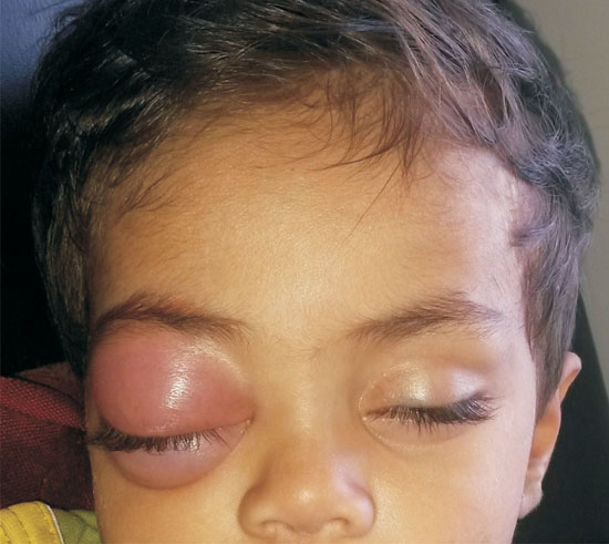
An 18-month-old toddler presented with fever and tender swelling of
right eye for 10 days (Fig. 1). We suspected orbital
cellulitis due to local signs of inflammation. Investigations were Hb 10
g/dL, platelet-count 336 x 10
3/L,
WBC 17.6 x 10⁹/L (85%
neutrophils), and C-reactive protein 56 mg/L. The puffiness of eye
reduced, though did not resolve completely following 5 days of
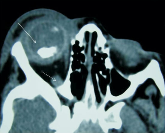
intravenous antibiotics. Cultures were sterile. CT demonstrated
calcified mass in the posterior segment of eye, with optic-nerve
thickening (Fig. 2). Examination under anesthesia
suggested retinoblastoma. CSF and bone marrow examination were
unremarkable. He received three cycles of chemotherapy, followed by
enucleation, further nine cycles of chemotherapy, and orbital
radiotherapy (stage III disease). Histopathology confirmed
retinoblastoma. Child is well one year following treatment.
 |
|
Fig. 1 Retinoblastoma presenting as orbital cellulitis
of the right eye in an 18-month-old boy.
|
 |
|
Fig. 2 CT scan showing intraocular
calcified mass in the right eye (solid arrow) with optic nerve
thickening (broken arrow).
|
Orbital cellulitis is an infrequent (4-5%)
presentation of retinoblastoma. Inflammation develops following tumor
necrosis. Fever, leucocytosis, anterior-segment extension, however not
necessarily extra-ocular spread, are characteristic. Prednisolone along
with antibiotics reduce inflammation, rendering definitive treatment
feasible. Presentation mimicking bacterial pre-septal cellulitis is
known in malignancies including retinoblastoma and rhabdomyosarcoma. A
calcified intraocular mass in such a case suggests the diagnosis of
retinoblastoma. Retrolental fibroplasia, Coats disease and toxoplasma
chorioretinitis can rarely develop calcification as late sequelae beyond
three years of age.
|
|
|
 |
|
