|
|
Letters to the Editor Indian Pediatrics 2005; 42:1167-1168 |
|||
|
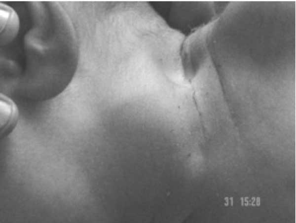
Bilateral Sternocleidomastoid Tumor in an Infant |
|||
|
Although, initial reports describe the right-sided lesion predominance, recent reports indicate no such predominance(3). The STOI is reported to be 32.7% in lower third of SCM and 43.3% in the middle third while the whole of the SCM is involved in 12.7% of cases(3). The tumor is more common in primipara and infants born with prolonged or difficult labor esp. breech deliveries. The STOI may be associated with torticollis (0.3% to 1.9%) and is usually associated with facial asymmetry and plagiocephaly (900%)(3). Co- existence of congenital hip dysplasia and other anomalies like mandibular hypoplasia, congenital spinal deformities, varus toes, metatarsus adductus, postural and structural equinovarus and calcaneal valgus have been reported. Bilateral neck masses in children can be diagnosed as benign tumor (STOI, lymph nodes) or malignant tumor (neuroblastoma, rhabdo-myosarcoma, fibrosarcoma). Although the diagnosis of the STOI is clinical, in doubtful cases with unusual presentation or if the tumor fails to resolve in 6 months time with conservative management, ultrasound, CT scan, MRI, FNAC and biopsy may help to confirm the diagnosis(1). Conservative management is recommended for STOI (success rate of 80-95%(3). The patients with rotational limitation upto 10º can be managed at home by exercises and positioning program taught to the parents. Manual stretching by professional physiotherapists is advised when the deficit of passive rotation is more than 10º. Complications of manual stretching include rupture of the SCM, bleeding or bruising (4.5%) and fractured clavicle (l.5%)(4). Surgery is indicated in patients with significant head tilt or deficit of passive rotation and the presence of tight band or tumor in the SCM, if they have not improved to acceptable levels after 6 months of manual stretching. Many surgical procedures described include simple open myotomy, extensive release of the SCM, bipolar release, myoplasties, or radical resections. The most important prognostic indicators for the surgical treatment are the severity of rotational deformity, clinical type and age of presentation(3). V. Manchanda,
|
![]()