1.gif (1892 bytes)

Images in Clinical Practice

Indian Pediatrics 2004; 41:-286

Sirenomelia


A 2-year-old female patient presented with complaint of gradual diminution of vision in the right eye for 3 months. It was associated with whitish discoloration of the pupillary region of the eye. There was no history of trauma, redness, swelling or discharge from the eyes. There was no past history of any fever or rashes. The family history was not significant. On clinical examination, the child could not perceive light in the right eye. The pupil in the right eye was dilated and not reacting to light. Fundus glow was absent on ophthalmoscopic examination. The child had leukocoria (while reflex) in the right eye (Fig. 1). A creamish white mass was seen filling almost the entire vitreous cavity behind the lens of the eye. The left eye was normal. The CT scan of orbits revealed the presence of an enlarged eyeball on the right side that harbored a solid mass in the posterior segment with multiple foci of calcification that was consistent with the diagnosis of retinoblastoma of the right eye.

Fig. 1. Leukocoria in the right eye.

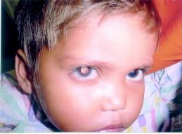
White pupillary light reflex (leukocoria) may indicate a disorder anywhere within the eye. Disorders include corneal opacity, blood (hyphaema) or other material in the anterior chamber, cataract, vitreous opacity or retinal disease. The most urgent diagnosis is retinoblastoma. Majority (70%) of cases of retinoblastoma present with leukocoria. Because it may be hereditary, a family history of retinoblastoma or of enucleation is of special concern. Although retinoblastoma is almost uniformly fatal without treatment, the cure rate is 90% or better when it is promptly recognized and treated and many children can be effectively treated without enucleation. Cataract can be congenital or acquired. Vitreous opacities present in persistent hyperplastic primary vitreous, organized vitreous hemorrhage, retrolental fibroplasias and endophthalmitis may present with leukocoria in childhood. Common retinal diseases that present with leukocoria include Coat’s disease, retinal dysplasia, retinal astrocytomas, retinal detachment and familial exudative vitreoretinopathy among others.

Harsh Gupta,
Pankaj Gupta,
Department of Pediatrics and Ophthalmology,
Vardhman Mahavir Medical College,
New Delhi 110 029,
India.
E-mail: [email protected]

Home

Past Issue

About IP

About IAP

Feedback

Links

 Author Info.

  Subscription