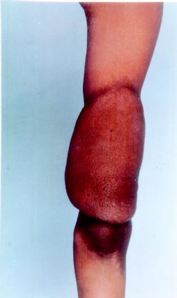
A 10-year-old boy presented with a gradually progressive swelling over
his right arm for last 7 years (Fig. 1). Examination revealed a
soft, diffuse swelling measuring 10 × 12 cm over posterior aspect of
right arm. The lesion was non-tender and freely mobile over the
underlying tissues. In addition, he had multiple hyperpigmented macules
with serrated margins over the trunk (cafe au lait macules) and
multiple freckle like macules (axillary freckles) in both axillae. He
also had multiple soft nodules in the skin (mollusca fibrosa) which were
widely dispersed over trunk and limbs. An ophthalmological examination
revealed multiple pigmented iris hamartomas (Lisch nodules) in both
eyes. A clinical diagnosis of Neurofibomatosis type I with plexiform
neurofibroma of right hand was made. A biopsy from lesion on right hand
showed a whorled proliferation of spindle shaped cells consistent with
neurofibroma.
|
 |
|
Fig. 1. Plexiform neurofibroma on right
elbow. |
Neurofibromatosis is a genodermatosis of
neuroectodermal origin characterized by multiple cutaneous turnours (mollusca
fibrosa), pigmented ‘cafe au lait’ macules, axillary freckles,
lisch nodules in iris and variable involvement of central nervous
system. The genetic defect is localised to chromosome 17 and is
transmitted in an autosomal dominant pattern. Plexiform neurofibroma
presents as a diffuse and elongated swelling along the course of a nerve
trunk/plexus, These tend to infiltrate into deeper structures like
fascia, muscles and bone. There is a localized or segmental hypertrophy
of underlying soft tissue resulting in a gross deformity of the involved
part. The incidence of malignant transformation into neurofibrosarcoma
is upto 5% of cases. Surgical excision is the treatment of choice,
Vijay Gandhi,
Subhav Aggarwal,
Department of Dermatology and STD,
UCMS and GTB Hospital,
Delhi, India.
