|
|
Personal Practice Indian Pediatrics 2003; 40:626-632 |
||||||||||||||||||||||||||||
|
Spirometry in Clinical Practice |
||||||||||||||||||||||||||||
|
From the Kanchi Kamakoti Childs Trust Hospital, Nungambakkam, Chennai 600 034.
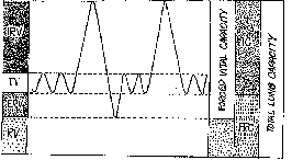
Table I

Lung Volumes and Capacities
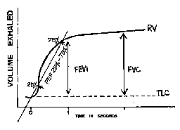
Spirometer Spirometers are instruments to conduct spirometry tests. Earlier, spirometers were of either rolling model or bellows type but the recent ones are electronic and fully automated using digital turbine and flow sensors that require no calibration. Though spirometry does not measure the individual lung volumes, it measures the forced vital capacity (FVC), which is a combination of tidal volume (TV) expiratory reserve volume (ERV) and inspiratory reserve volume (IRV). The other indices like forced expiratory volume in one second (FEV1), the ratio of FEV1 with FVC (FEV1/FVC), forced expiratory flow 25% to 75% of forced vital capacity (FEF 25%-75%) are measured from FVC. The Spirometer cannot measure functional residual capacity (FRC) or total lung capacity (TLC) but these parameters are not important in routine assess-ment of common lung problems. Baseline spirometric values depend on various factors like race, sex, age, etc. The standing height is a satisfactory predictor of lung function(4). Spirometry Test Children above 5 to 6 years of age can produce an acceptable FVC curve with adequate coaching. The environment for testing should be child friendly. Before attempting spirometry, it is important to make the child familiar with the laboratory, instruments and persons. The mouthpiece is given 1-2 days in advance for the child to practice at home, so that he/she will be comfortable and confident to perform the procedure that will definitely help in obtaining better results. The child’s torso and head should be erect during the procedure. Application of nose clips may yield better results. To achieve a good FVC the child should take a slow breath to full inhalation followed by a brief hold and then a sustained exhalation with maximum effort without coughing or quitting during the procedure. The child should be coached and encouraged during expiration to achieve a complete forced vital capacity i.e., "blowing" as long as possible for at least 3 seconds. FVC manoeuvre in spirometry is like that of balloon blowing and this example will make understanding of the technique easy. The same person, preferably a doctor should perform and interpret spirometry. An ideal spirometry should include at least two reproducible curves with a difference of less than 5% and the best accepted curve is the one that has the largest sum of FEV1 and FVC(5). The child is allowed more than three attempts to achieve the above. The spirometer usually computes the largest value of FEV1 and FVC even if these values are from two different curves(6). Forced vital capacity is the difference between total lung capacity and residual volume and is generated by maximum expiration after maximum inspiration. Normally it is reached within 3 to 4 seconds. The most important aspect of spirometry is to produce a good forced vital capacity curve for the specified period without quitting or coughing. During the FVC maneuver the expiratory volume is plotted against time and it is called as the time volume curve (Fig 2). Spirometry indices are reported comparing the individual’s value along with the predicted values. In normal individuals more than 80% FVC can be achieved in the first one second. The FEV1 denotes the fraction of forced vital capacity expired during the first second. The ratio of FEV1 to FVC is usually referred as forced expiratory ratio. Forced expiratory flow 25-75% is measured from FVC curve by excluding first 25% and last 25% of expiratory flow (FEF 25-75%) and sometimes referred as maximal mid expiratory flow rate (MMFR-25-75%), this mostly evaluates the small airways.
FVC will be diminished in both obstructive and restrictive diseases. In the early stages of obstruction, FVC may slightly increase due to air trapping. If the child quits before the end of the FVC manoeuvre, the FVC is underestimated. Consequently the FEV1/FVC ratio may be normal resulting in a wrong interpretation as restrictive lung disease instead of obstructive disease. FEV1 signals airway obstruction. The FEV1/FVC ratio is decreased in obstructive diseases because the rate of airflow is slowed. In restrictive lung diseases, it is normal or even higher than normal because both FVC and FEV1 are reduced proportionally. FEF 25-75% is a more sensitive indicator of small airway obstruction than FEV1. In early or mild asthmatics because of air trapping, TLC will increase but FEV1 and FEV1/FVC ratio is deceptively normal. In such conditions, the measurement of FEF 25-75% may be diagnostic. From FVC curve several indices can be derived which may be confusing for the beginner. Concentrating on the above discussed basic spirometry indices will fetch more than 75% of relevant information. The important indices that categorize the lung disease into obstructive and restrictive types are FVC, FEV1, FEV1/FVC ratio and FEF 25-75% (Table II). Table II Spirometry Indices in Respiratory Diseases.
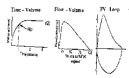
Modern Spirometry Curves Time volume curves are conventionally used to measure forced vital capacity in all spirometers. Spirometry is a growing field and the forced vital capacity curves are also getting metamorphosed. In addition to time-volume curve, modern spirometry machine presents new plots like flow volume curve and flow volume loops (Fig 3). In a time-volume curve the time is plotted against volume, whereas if the expiratory flow is plotted against lung volume it is called as Flow volume curve which is another way of visualizing the time volume curve. In a flow volume loop the expiratory flow rate is recorded against the expired volume. In this study, the patient performs the FVC manoeuvre and at completion, he or she is requested to perform full inspiration. Though the new curves do not add any additional information, graphic illustration gives a quick assessment of the disease pattern. Since all the modern spirometers depict flow volume loops, knowledge about flow volume loop is mandatory for the interpretation of modern spirometry.
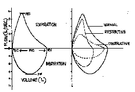
Fig. 3. Depiction of spirometry curves in different methods. Whereas in time volume and flow volume curves expiration is measured, in flow volume loop both expiration (upper half) and inspiration (lower half) are measured. Flow Volume Loop In flow volume loop, upper half of the curve represents expiration while the lower half inspiration, forming a loop and the direction of the loops is clockwise(7) (Fig. 4). Reduction of vertical axis (Y-axis) represents reduction of flow representing obstructive lung problems. Reduction of horizontal axis (X-axis) denotes reduction of lung volume thereby indicating restrictive lung diseases. Obstructive lung disease alters the shape of the loop while restrictive lung disease alters the size of the loop. Thus, flow volume loop provides a graphic picture to classify pulmonary disease and to locate the site of obstruction. In restrictive lung disease, all lung volumes and flows (inspiratory and expiratory) are reduced resulting in a small loop without any change in the shape (Fig. 4).
Fig. 4. Normal flow volume curve and curves in different pulmonary conditions. Intrathoracic obstruction reduces all expiratory flows and as the flow reduction becomes more severe, there is concavity or "scoop" in the expiratory limb of loop. In distal obstruction (e.g., asthma) flows are most affected at low volumes and in proximal obstruction (e.g., tracheal pathology) flows are affected at high volumes. In extra thoracic obstruction there is flattening of the inspiratory limb of the loop (lower portion of the loop) without altering the expiratory limb. In clinical conditions with mixed airway obstruction, the ratio of maximum expiratory flow (Vmax50%) to maximum inspiratory flow (MIF 50%) is used. In normal individual, the ratio is equal to 1. (Vmax 50%/MIF 50% = 1). In variable intra thoracic obstruction the ratio is <1 and in variable extra thoracic obstruction the ratio is >1. Spirometry Indications The indications of spirometry have been increasing over the years. Though etiological diagnosis is not possible it is used to assess the functional derangement in many lung diseases. Spirometry plays a significant role in the management of many chronic lung conditions like asthma, chronic obstructive airway disease (COPD), and interstitial lung disease and as a preoperative assessment before cardiopulmonary surgery. Lung Disease The most important indication for spirometry is to differentiate lung diseases into obstructive and restrictive for effective management. The common obstructive lung disease in children is asthma and other obstructive disorders are bronchiectasis, cystic fibrosis, chronic obstructive airway disease (COPD). Restrictive lung diseases include structural diseases of the chest wall (kyphoscoliosis), neuromuscular problems and interstitial lung diseases (ILD). Obstructive diseases are characterized by low flows with near normal volumes whereas restrictive lung diseases by small volumes and normal flows. Asthma The urgent issue in asthma management strategy is early diagnosis. Under diagnosis of asthma leads to under treatment that causes progressive remodeling in the airway mucosa. Recent studies have demonstrated (on the basis of broncho alveolar lavage) inflam-matory changes even in mild persistent asthma(8). Though asthma can be diagnosed on clinical grounds, poor compliance and difficulty in monitoring are the impedence in successful management. Recent studies indicate that upto 70% of patients with asthma do not comply with treatment(9). So all older children should be subjected to spirometry in the initial evaluation of the disease. In the majority, the demonstration of the objective deviation of the observed value (>20%) from the predicted value by spirometer confirms asthma. When spirometry values are normal and asthma is strongly suspected the response to bronchodilator aerosol is measured (Bronchodilator challenge test). Reversible airway obstruction characterized by a rise in the FEV1 and/or FVC by atleast 12% (from pre to post bronchodilator), is characteristic of asthma(10). Airway obstruction due to fixed anatomic obstruction may not respond. Most of the asthmatics exhibit exercise intolerance and this is used for diagnosis of asthma (Exercise testing). Child is made to excercise for 6 minutes. Tread mill or bicycle ergometer are preferred methods of testing in children to provide exercise-induced broncho-spasm to diagnose asthma. Flow rates should be measured 3,10 and 15 minutes following exercise. A drop in FEV1 of 10% or more is taken as positive test(11). Measuring other parameters of obstruction such as FEF 25-75% of FVC and PEFR increases the sensitivity of the test. Though spirometry forms the cornerstone of asthma diagnosis, some individuals with asthma may have near normal spirometry and may not show significant bronchodilator response. In such cases challenge testing with inhaled histamine or methacholine may help to make a diagnosis before starting empirical therapy. Increasing dose of histamine (0.06, 0.12, 1.00, 2.50 mg/mL) or methacholine (25 mg/mL) is administered and FEV1 measured before and after the test. A 20% decrease in FEV1 when compared to the baseline value is a positive response. However, in routine practice, challenge tests are rarely required. In addition to diagnosis, comparison of the curves/loops during therapy will evoke interest among parents and involve them in the partnership management of asthma. Peak flow meters and asthma diary may further increase patient compliance. Chronic obstructive pulmonary disease (COPD) Adolescents and older children addicted to the habit of smoking, those who are exposed to high environmental pollution or bio fuel smoke are potential candidates of COPD. Since clinical presentation of COPD is invariably cough, the majority may ignore this problem for a long period of time. Subjecting this group for early spirometry will alert them about this progressively fatal disease. In COPD unlike in asthma, bronchodilator reversibility test may be negative and FEV1 may progressively fall. Interstitial lung disease (ILD) Interstitial lung disease is a heterogenous group of disorders of varied etiology with common clinicoradiological presentation. Since majority present with cough and exertional dyspnea they will be treated as asthma or tuberculosis for prolonged periods. Though lung biopsy is the confirmatory test, spirometry plays a significant role in the diagnosis and monitoring the course of the diasese. Restrictive spirometry pattern is typically observed in ILD due to reduction of the static lung volumes. Reduction of FVC is greater than FEV1 resulting in normal or supernatural FEV1/FVC ratio(12). Lung surgery All children planned for lobectomy or pneumonectomy require spirometry tests to assess whether there will be adequate lung sufficiency after lung resection. Children contemplating for pneumonectomy should have FVC of more than 2 liters. Preoperative work up Spirometry plays a major role in the preoperative work up of structural anomalies of chest wall diseases. If the preoperative inspiratory capacity is less than 30 mL/kg the child may require assisted ventilation in the postoperative period. Peak Flow Meter Though Spirometry is a gold standard test to diagnose asthma, peak (expiratory) flow meter (PEF) can be a useful alternative to predict underlying asthma when spirometry is not available. Peak flow meter records peak expiratory flow rate (PEFR). Peak expiratory flow rate is the greatest flow obtained on forced expiration after complete inspiration using Peak flow meter. Peak flow rates are effort dependent and measure mostly large airway function. Though PEFR correlate well with FEV1, it is not a substitute for spirometry. A difference of 20% or more between morning and night values is considered a good predictor of asthma. Since early asthma can be missed by PEFR measurement, spirometry should be preferred to diagnose asthma. However, PEFR plays a major role in asthma follow up. A sudden fall of PEFR may be an early warning of impending attack of asthma(2). PEFR plays a major role to monitor asthma therapy and serial recording will reflect the prognosis of the disease as well as outcome of therapy. Further, it provides an objective assessment of lung function in those children with asthma who are unable to do a FVC procedure. Respiratory diseases are at an upsurge globally. Early objective evidence of pulmonary disease will ensure adequate compliance and successful management. In the era of consumerism, evidence based approach is in demand and spirometers play a significant role in management strategy of many respiratory illnesses, especially asthma. Basic Knowledge about PFT will help practitioners in early identification of respiratory problem and its successful management. Contributors: DV and LS conceived and designed the article. AB and SS drafted the manuscript. All the authors were involved in literature search and final approval. Funding: None. Competing interest: None stated.
| ||||||||||||||||||||||||||||
|
References | ||||||||||||||||||||||||||||
|
|
![]()